Showing posts with label Antibacterial. Show all posts

Sea Urchin Methanolic Extract Shows Antibacterial Activity Against E. coli and S. aureus | InformativeBD

Pharmacological activity of the methanolic extract of sea urchins against Escherichia coli and Staphylococcus aureus

Kate Jocel D. Barroga, Diana C. Castillo, and Evaristo A. Abella, from the different institute of Philippines. wrote a Reseach Article about, Sea Urchin Methanolic Extract Shows Antibacterial Activity Against E. coli and S. aureus. Entitled, Pharmacological activity of the methanolic extract of sea urchins against Escherichia coli and Staphylococcus aureus. This research paper published by the International Journal of Biosciences (IJB). an open access scholarly research journal on Biosciences. under the affiliation of the International Network For Natural Sciences| INNSpub. an open access multidisciplinary research journal publisher.

Abstract

This study elucidated the pharmacological potential of sea urchins using methanol as extracting medium. The antibacterial potential was evaluated using the paper disc method and zone of inhibition against Escherichia coli and Staphylococcus aureus was measured. Antioxidant properties of sea urchins were evaluated using DPPH radical scavenging assay. Three species of sea urchin randomly collected along the intertidal zone of Diguisit, Baler Aurora were identified using diagnostic keys by the National Museum of the Philippines and they were identified as follows; Echinothrix diadema, Echinometra mathaei, and Echinometra oblonga. E. diadema recorded the highest diameter zone of inhibition against E. coli and S. aureus after 24 hours of incubation with 11.03 ± 1.75mm and 13.52 ± 1.13mm respectively while E. mathaei only inhibited S. aureus with zone of inhibition of 9.27 ± 2.06mm in 24 hours of incubation as well. As the zone of inhibition prolongs, the zone of inhibition decreases as observed in 48 hours of incubation. E. oblonga did not show inhibitoy effect, however it recorded the highest radical scavenging activity with 64.46% among the three species of sea urchins. This was followed by E. mathaei (51.52%) and E. diadema (37.38%). All collected species manifested antioxidant potential. Based on the results, the collected species of sea urchins has a pharmacological potential.

Submit your article to IJB Journal

Read more Mapping Dindéresso Forest Landscapes with Sentinel-2 and Machine Learning | InformativeBD

Introduction

Marine invertebrates are excellent sources of bioactive compounds with antibacterial and antioxidant dynamics. Recent discovery on pharmacological dynamics has stimulated the search for natural agents or natural sources that will lead to a desirable antibacterial and secondary metabolites (Abubakar et al., 2012). 

Pharmacological activity of the methanolic extract of sea urchins against Escherichia coli and Staphylococcus aureus

The diversity of marine organisms in our ecosystem, secondary metabolites has been identified as one of very important compound available produced in marine organisms. Sea urchins are small, spiny, globular animals belong to the class Echinoidea of the echinoderm phylum (Shankarlal et al., 2011). Additionally, sea urchins have orbicular bodies coated with a strict shell and thoroughly covered with many sharp spines (Amarowicz et al., 2012).

As stated in the study of Bich et al., (2004), echinoderms have pharmacologically active secondary metabolites. The antibacterial activity of sea urchin is generally assayed through various extracts with different solvents. Methanol extract of Tripneustes gratilla showed highest antimicrobial activity against Pseudomonas aeruginosa (Abubakar et al., 2012). Also, methanol extract of Diadema setosum exhibited higher zone of inhibition against Salmonella typhimurium, Staphylococcus epidermidis, Citrobacter freundii, and Klebsiella pneumoniae (Rahman et al., 2015). 

Pharmacological activity of the methanolic extract of sea urchins against Escherichia coli and Staphylococcus aureus

An investigation report of Bragadeeswaran et al. (2013) on the bioactive compounds of sea urchin Temnopleurus toreumaticus showed remarkable hemolytic and cytotoxic activities. The spines of purple sea urchin Strongylocentrotus nudus showed excellent activity by using DPPH scavenging activity indicating the presence of PHNQ as potential sources of natural antioxidants (Zhou et al., 2011). 

The present work focused on the screening of the antibacterial and antioxidant activity of whole globular body and tissues of Echinothrix diadema, Echinometra mathaei, and Echinometra oblonga collected from the intertidal zone of the coastal ecosystem of Barangay Diguisit, Baler, Aurora, Philippines.

Reference

Abubakar LA, Mwangi CM, Uku JU, Ndirangu SN. 2011. Antimicrobial activity of various extracts of the sea urchin Tripneustes gratilla (Echinoidea). African Journal of Pharmacology and Therapeutics 1(1).

Amarowicz R, Synowiecki J, Shahidi F. 2012. Chemical composition of shells from red (Strongylocentrotus franciscanus) and green (Strongylocentrotus droebachiensis) sea urchin. Journal of Food Chemistry 133, 822-826.

Bich D, Chung D, Chuong B, Dong N, Dam D, Hien P, Lo V, Mai P, Man P, Nhu D. 2004. The medicinal plants and animals in Vietnam. Hanoi Science and Technology Publication 1.

Bragadeeswaran S, Sri Kumaran N, Prasath Sankar P, Prabahar R. 2013. Bioactive potential of sea urchin Temnopleurus toreumaticus from Devanampattinam, Southeast coast of India. Journal of Pharmacy and Alternative Medicine 2(3), 9-18.

Brasseur L, Hennebert E, Fievez L, Caulier G, Bureau F, Tafforeau L, Eeckhaut I. 2017. The roles of spinochromes in four shallow water tropical sea urchins and their potential as bioactive pharmacological agents. Marine drugs 15(6), 179.

Horton, J. 2012. Invertebrates of the Coral Sea:  Echinometra mathaei. Published at the University of Queensland, Australia.

Kazemi S. Heidari B, Rassa M. 2016. Antibacterial and hemolytic effects of aqueous and organic extracts from different tissues of sea urchin Echinometra mathaei on pathogenic streptococci. International Aquatatic Research 8(4), 299-308.

Lalitha, M. K. 2004. Manual on Antimicrobial Susceptibility Testing. New Delhi, India. Indian Association of Medical Microbiologists.

Lebedev AV, Ivanova MV, Levitsky DO. 2003. Iron chelators and free radical scavengers in naturally occurring polyhydroxylated 1, 4-naphthoquinones. Hemoglobin 32(1-2), 165-179.

Minh CV, Kiem PV, Huong LM, Kim YH. 2004. Cytotoxic constituents of Diadema setosum. Archives of Pharmacal Research 27(7), 734-737.

Mokhlesi A, Saeidnia S, Gohari AR, Shahverdi AR, Nasrolahi A, Farahani F, Es’haghi N. 2012. Biological activities of the sea cucumber Holothuria leucospilota. Asian Journal of Animal and Veterinary Advances 7, 243-249.

Pettit GR, Ode RH. 1979. Phenols, quinones, and related compounds. Phenols, quinones, and related compounds. In: Biosynthetic Products for Cancer Chemotherapy.

Rahman MA, Amin SMN, Yussof FM, Kupan P, Sham-Sudi MN. 2012. Length weight relationships and fecun-dity estimates of long-spined sea urchin, Diadema setosum, fron Pulau Pangkor, Peninsular Malaysia. Aquatic Ecosystem Health Manage 15, 311-315.

Schoppe S. 2001. Echinoderms of the Philippines: A guide to common shallow water sea stars, brittle stars, sea urchins, sea cucumbers and feather star. VISCA-GTZ Program on Applied Tropical Ecology, Visayas State College of Agriculture, Leyte, Philippines.

Shankarlal S, Prabu K, Natarajan E. 2011. Antimicrobial and antioxidant activity of purple sea urchin shell (Salmacis virgulata L. Agassiz and Desor 1846). American-Eurasian Journal of Science Research 6(3), 178-181.

Silhavy TJ, Kahne D, Walker S. 2010. The bacterial cell envelope. Biosynthetic Products for Cancer Chemotherapy 127-143.

Yasmin. 2015. Developmental biology of sea urchin, Echinometra oblonga (Blainville, 1825) And Its Larval Settlement Behavior in Response to Chemical Cues.

Zhou DY, Zhu BW, Wang XD, Tan H, Yang J, Li DM, Dong XP, Wu HT, Sun L, Li XL, Murata Y. 2011. Extraction and antioxidant property of polyhydroxylated naphthoquinone pigments from spines of purple sea urchin Strongylocentrotus nudus. Food Chemistry (129), 1591-1597.

SourcePharmacological activity of the methanolic extract of sea urchins against Escherichia coli and Staphylococcus aureus  

Maranta Arundinacea: Antibacterial Extracts Study | InformativeBD

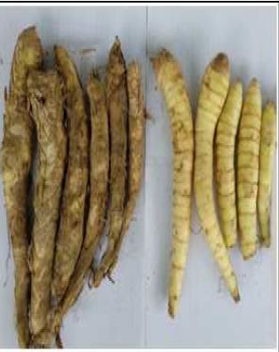
In vitro antibacterial effect of the extracts of Maranta arundinacea rhizomes against selected pathogens

Balan Aswini, Sivagurunathan Paramasivam, Sivaprakasam Sowndarya and Uma Chinnaiyan from the different institute of the India. wrote a research article about, Maranta Arundinacea: Antibacterial Extracts Study, entitled,  "In vitro antibacterial effect of the extracts of Maranta arundinacea rhizomes against selected pathogens". This research paper published by the International Journal of Biosciences | IJB . an open access scholarly research journal on Biosciences under the affiliation of the International Network For Natural Sciences | INNSpub. an open access multidisciplinary research journal publisher.

Abstract

Plants are known to synthesize an array of secondary metabolites referred to as phytochemicals that have disease-prevention properties. Potential efficacy and minimum to no side effects are key advantages of plant-derived products, making them sustainable choices for medical treatments. The aim is to investigate the antibacterial activity and phytochemical screening of methanolic, ethanolic, ethyl acetate and chloroform extracts of Maranta arundinacea (arrowroot) rhizomes. New antimicrobial agents need to be developed to battle the rapidly evolving pathogens.

Submit your article to IJB Journal

Read moreCellulase Overproduction: Trichoderma harzianum Mutants | InformativeBD

Introduction

The emergence and spread of antibiotic resistance, as well as the evolution of new strains of disease-causing agents, are of great concern to the global health community. Effective treatment of a disease entails the development of new pharmaceuticals or some potential source of novel drugs. Commonly used medicinal plants of our community could be an excellent source of drugs to fight off this problem (Manandhar et al., 2019). The vast number of medicinal plants have been recognized as valuable resources of natural antimicrobial compounds as an alternative that can potentially be effective in the treatment of these problematic bacterial infections (Iwu et al., 1999). According to the World Health Organization (WHO), medicinal plants would be the best source to obtain a variety of drugs (WHO, 2002).

UTIs are contagious infection, affecting both the male and female population and can infect various parts of the urinary tract system (UTS), such as urethra, ureter, urinary bladder and kidneys mainly focused on the lower UTS like urethra and ureter.

Escherichia coli and Klebsiella sp are the main causative agents, although other Gram-positive bacteria and even fungi have also been isolated in numerous infected population (Flores-Mireles et al., 2015). Herbal formulation involves the use of fresh or dried plant parts. The exact mechanisms of medicinal herbs and their phytochemical constituents that are responsible for the effect on UTI are still to be investigated. Further research is needed to elucidate clearly the mode of action of these phytochemicals. Additional studies are needed to confirm the phytoconstituents that are responsible for the treatment of UTI (Aswini et al., 2022).

The arrowroot plant M. arundinacea L. is identified to possess phytochemicals that make them medically important in exhibiting antidiarrheal, probiotic, antiulcer, antioxidant, antimicrobial, vibriocidal and immunostimulatory effects (Firoskhan and Muthuswamy, 2021). Considering the vast potentiality of plants as sources for antimicrobial drugs, this study aimed to investigate invitro antibacterial activity of extracts from some selected medicinal plants from Nepal against the most common microbial pathogens including MDR (Multi- Drug Resistant) bacteria (Manandhar et al., 2019).

The aim of this research is to study different extracts for its antimicrobial activity and phytochemical screening. It is a preliminary step for the identification of medicinal value of the plant.

Reference

Ahmed el-HM, Nour BY, Mohammed YG, Khalid HS. 2010. Antiplasmodial activity of some medicinal plants used in Sudanese folk-medicine. Environmental Health Insights 4(4), 1-6.

Aswini B, Sivagurunathan P, Sowndarya S, Sumathi V, Vivekanandhan S, Uma C. 2022. A Study On The Effect Of Methanolic Extracts Of Maranta Arundinacea Leaves Against Urinary Tract Infection Causing Pathogens. Uttar Pradesh Journal of Zoology 43(22), 71–77. https://doi.org/10.56557/UPJOZ/2022/v43i223235

Elumalai EK, Ramachandran M, Thirumalai T, Vinothkumar P. 2011. Antibacterial activity of various leaf extracts of Merremia emarginata. Asian Pacific Journal of Tropical Biomedicine 1(5), 406–408. https://doi.org/10.1016/S2221-1691(11)60089-0

Firoskhan N, Muthuswamy R. 2021. Review on Maranta arundinacea L. (Marantacea) International Journal of Pharmacognosy and Pharmaceutical Research. International Journal of Pharmacognosy and Pharmaceutical Research Genus 3(1), 1–4.

Jayakumar A, dan Suganthi A. 2017 “Biochemical and phytochemical analysis of Maranta arundinacea (L.) Rhizome,” International Journal of Research in Pharmacy and Pharmaceutical Sciences, 2(3), hal. 26–30.

Julianeli TDL, Jackson RGS, Kelly S, Ana Sílvia SC.  2011. Selective spasmolytic effect of a new furanoflavoquinone derivative from diplotropin on guinea-pig trachea. Journal of Chemical and   Pharmaceutical Research 3(1), 249-258.

Kudo YH, Kobayashi A, Konishi YS, Kondo K. 2004. Antibacterial activity of plants used in cooking aroma and taste. Journal of Food Protocol 67, 2820-2824.

Iwu MW Duncan AR, Okunji CO. 1999. “New antimicrobials of plant origin in. Perspectives on new crops and new uses,” in Plant Breeding Reviews, J. Janick, Ed., ASHS Press, Alexandria, Virginia.

Maria Lysete AB, Maria Raquel FL. 2009. Studies on the antimicrobial activity and brine shrimp toxicity of Z. tuberculosa extracts and their main constituents. Annals of Clil Microb Antimic 8, 16.

Manandhar S, Luitel S, Dahal RK. 2019. In Vitro Antimicrobial Activity of Some Medicinal Plants against Human Pathogenic Bacteria. Journal of Tropical Medicine. https://doi.org/10.1155/2019/1895340

Mehrangiz KK, Seyed AE, Masoud SG, Esmaeel AS, Amirhossein S. 2011. Antiviral activities of aerial subsets of Artemisia species against Herpes Simplex virus type 1 (HSV1) in vitro. Asian Biomed 5(1), 63-68.

Mohamed Sham Shihabudeen H, Hansi Priscilla D, Kavitha T. 2010. Antimicrobial activity and phytochemical analysis of selected Indian folk medicinal plants. International Journal of Pharmaceutical Sciences and Research 1(10), 430-434.

Pérez E, Lares M. 2005. Plant Foods for Human Nutrition 60(3), 113–116. https://doi.org/10.1007/s11130-005-6838-9

Rahman MK, Chowdhury MAU, Islam MT, Chowdhury MA, Uddin ME, Sumi CD. 2015. Evaluation of Antidiarrheal Activity of Methanolic Extract of Maranta arundinacea Linn. Leaves. Advances in Pharmacological Sciences 1–6.

Rahman MK, Chowdhury MAU, Islam MT, Chowdhury MA, Uddin ME, Sumi CD. 2015. Evaluation of Antidiarrheal Activity of Methanolic Extract of Maranta arundinacea Linn. Leaves. Advances in Pharmacological Sciences. https://doi.org/10.1155/2015/257057

Rajashekhara N, Ashok B, Sharma P, Ravishankar B. 2014. (An International Quarterly Journal of Research in Ayurveda) 35(4), 433. https://doi.org10.4103/0974-8520.159013

Ren L. 2014. “Antibacterial Activities of Flavonoids: Structure Activity Relationship and Mechanism,” Current Medicinal Chemistry 22(1), hal. 132–149. https://doi.org/10.2174/0929867321666140916113443

Shan B, Cai YZ, Brooks JD, Corke H. 2007. The in vitro antibacterial activity of dietery spices and medicinal herbs extracts. International Journal of Microbiology 117, 112-119.

Snathi R, Lakshmi G, Priyadharshini AM, Anandraraj C. 2011. Phytoscreening of Nerium ollender leaves and M. charatia leaves. International research journal of pharmacy 2(1), 131-135.

Sholichah E, Deswina P, Sarifudin A, Andriansyah CE, Rahman N. 2019. Physicochemical, structural and morphological properties of some arrowroot (Maranta arundinacea) accessions growth in Indonesia. AIP Conference Proceedings 2175. https://doi.org/10.1063/1.5134572

World Health Organization (WHO). 2002. Traditional Medicine Strategy, Geneva.

Wu C, Chen F, Wang X, Kim HJ, He GQ, Haley-Zitlin V, Huang G. 2006. Antioxidant constituents in feverfew (Tanacetum parthenium) extract and their chromatographic quantification. Food chemistry 96, 220-227.

SourceIn vitro antibacterial effect of the extracts of Maranta arundinacea rhizomes against selected pathogens