Shea Caterpillar Flour in Tilapia Feed: Impact on Growth Performance | InformativeBD

Effects of incorporation of shea caterpillar flour (Cirina butyrospermi) in the feed on the growth performance of tilapia Oreochromis niloticus (Linnaeus, 1758) raised in basin brazil strain

Kolotcholoman Silue,  from the institute of Ivory Coast . N'golo Ouattara, from the institute of Ivory Coast. Medard Gbai, from the institute of Ivory Coast. and Kouakou Yao, from the institute of Ivory Coast. wrote a Research Article about, Shea Caterpillar Flour in Tilapia Feed: Impact on Growth Performance. Entitled, Effects of incorporation of shea caterpillar flour (Cirina butyrospermi) in the feed on the growth performance of tilapia Oreochromis niloticus (Linnaeus, 1758) raised in basin brazil strain. This research paper published by the International Journal of Biosciences (IJB)an open access scholarly research journal on Biosciences . under the affiliation of the International Network For Natural Sciences| INNSpub. an open access multidisciplinary research journal publisher.

Abstract

With the aim of seeking alternatives to the expensive use of fish meal in fish feed, a study was carried out on the incorporation of shea caterpillar meal (Cirina butyrospermi) into the diet of Oreochromis niloticus. Three diets with progressive incorporation rates of 15% (A15), 20% (A20) and 25% (A25) of C. butyrospermi meal were prepared from a local feed based on agricultural by-products and compared with a control diet (AT) without animal meal. Tilapia larvae with a mean initial weight of 4± 0.05 g were reared in duplicate basins at a stocking density of 100 fry per m3. The fish were fed 4 times a day with a ration of 5% of their total biomass from the start to the end of the experiment. After 90 days of rearing, the best feed conversion and final weight (1.50 ± 0.14; 37.10± 2.25 g, respectively) were obtained with diet A20, followed by diet A15 (1.62 ± 0.08; 35.20± 2.86 g) and A25 (1.74 ± 0.12; 33.03± 3.44 g). The lowest daily growth (0.33± 0.03 g/day) and the highest feed conversion index (1.99± 0.12) were recorded with the AT diet. At the end of the experiment, survival rates were greater than or equal to 92.86%. At the end of this study, shea caterpillar meal can be incorporated into tilapia feed and considered a good protein source.

Submit your article to IJB Journal

Read more Impact of Rooting Hormones on Survival of Three Bamboo Species | InformativeBD

Introduction 

To date, over 80% of global fish production depends on exogenous feed in the form of supplements to feed from the farming environment (FAO, 2018). Several investigations have been carried out in the field of feed, contributing to the rapid expansion and spectacular development of aquaculture worldwide (Kolditz, 2008; FAO, 2018). Against this backdrop, we are witnessing an intensification of fish farming, with widespread and significant use of artificial feeds (Gaye-Siessegger et al., 2005). These highperformance feeds, which provide fish with a complete diet, are produced in developed countries. However, in developing countries, the difficulty lies in acquiring suitable exogenous feeds for feeding fish, especially at juvenile stages (Hung et al., 2001; Coulibaly et al., 2007; Pangni et al., 2008).

These exogenous feeds are considered to be very expensive and, above all, difficult to access, which hampers the expansion of the fisheries sector (Barrows and Hardy, 2001).

Effects of incorporation of shea caterpillar flour (Cirina butyrospermi) in the feed on the growth performance of tilapia Oreochromis niloticus (Linnaeus, 1758) raised in basin brazil strain

The high cost of these feeds is most often due to the use of fishmeal as a protein source. Indeed, the extensive use of fishmeal in feeds is at the root of this high cost (Nguyen et al., 2009; Hardy, 2010). These authors also point out that this input is becoming increasingly scarce and expensive, representing a major limiting factor to aquaculture developments. Faced with this situation, the use of adapted local feeds is necessary to maximize feed conversion efficiency and growth (Guzel and Arvas, 2011).

In Côte d'Ivoire, for example, Oreochromis niloticus production faces several obstacles. Among these obstacles are (i) the low nutritional quality of local feed (ii) the low availability of quality imported feed (iii) the cost of imported feed deemed high by stakeholders, and above all (iv) the lack of fry (MIRAH, 2014, PREPICO, 2019). Yet it has been reported by several authors (Richter et al., 2003; Liebert and Portz, 2005; Zhao et al., 2010) that in tilapia, combining several agricultural by-products with insect meal in feed formulas can reduce production costs while improving fish growth. However, in Côte d'Ivoire, there is a wide range of agricultural by-products that have already been tested in fish feeds (Nguyen et al., 2009; Zhao et al., 2010; Bamba et al., 2018). These include cottonseed and soybean meal, and rice and wheat bran. These raw materials are found in abundant quantities in Côte d'Ivoire (Sangare et al., 2009; FAO, 2014). For all these aforementioned reasons, we chose to use compound feeds based on agricultural by-products and shea caterpillar (C. butyrospermi) meal in the diet of pond-reared fry. With this in mind, the general objective of this study was to test the use of shea caterpillar meal (C. butyrospermi) combined with local agricultural by-products in the diets of tilapia Oreochromis niloticus "Brazil strain" fry.

Reference

Azaza MS, Mensi F, Abdelmouleh A, Kraïem MM. 2005. Development of dry feeds for Nile tilapia Oreochromis niloticus (L., 1758) reared in geothermal waters of southern Tunisia. Bulletin de l’Institut National des Sciences et Technologies de la Mer de Salammbô 32, 23–30.

Balarin JD, Haller RD. 1982. The intensive culture of tilapia in tanks, raceways and cages. In: Muir JF, Roberts RJ, editors. Recent Advances in Aquaculture, Vol. 1. Croom Helm, London, 265–356.

Bamba Y, Ouattara A, Moreau J, Gourene G. 2007. Relative intake of natural and artificial food in the diet of tilapia Oreochromis niloticus in captivity. Bulletin Français de la Pêche et de la Pisciculture 386, 55–68.

Bamba Y, Ouattara N, Ouattara S, Ouattara A, Gourene G. 2014. Effect of diets containing cocoa bean shell and coconut oil cake on the growth of Oreochromis niloticus (Linne, 1758) in pond. International Journal of Biological and Chemical Sciences 8, 1368–1380.

Bamba Y, Ouattara S, Ouattara N, Doumbia L, Ouattara A, DA Costa KS, Gourene G. 2018. Effects of feeding different plant protein sources in combination with cocoa peel, peanut skin, and copra cake on growth performance of tilapia Oreochromis niloticus (Linnaeus, 1758). Science et Technique, Sciences Naturelles et Appliquées. Special Issue 4, 531–544.

Barrows FT, Hardy RW. 2001. Nutrition and feeding. In: Wedemeyer GA, editor. Fish Hatchery Management (2nd ed.). John Wiley & Sons, New York, NY, USA, 483–558.

Campbell D. 1978. Formulation of feeds for rearing Tilapia nilotica in Lac de Kossou, Côte d’Ivoire, 31.

Coulibaly A, Ouattara NI, Kone T, N’Douba V, Snoeks J, Goore BG, Kouamelan EP. 2007. First results of floating cage culture of the African catfish Heterobranchus longifilis Valenciennes, 1840: Effect of stocking density on survival and growth rates. Aquaculture 263, 61–67.

Coulibaly ND, Kondombo C, Imien HL. 2021. Effects of partial substitution of fish meal by maggot meal on the growth of Nile tilapia (Oreochromis niloticus Linnaeus, 1758). Journal of Animal and Plant Sciences 50, 50–61.

Diabagate Y, Bamba Y, Brou KJL, N’zue KR, Ouattara A. 2024. Effects of local feeds containing copra and cotton cakes on pond production performance of juvenile tilapia Oreochromis niloticus (Linnaeus, 1758) “Brazil strain”. Sciences de la Vie, de la Terre et Agronomie 11, 1249–1256.

Dibala CI, Yougbaré MC, Konaté K, Coulibaly ND, Dicko MH. 2018. Production of Nile tilapia (Oreochromis niloticus Linneaus, 1758) with plant protein-based feeds. Journal of Applied Biosciences 128, 12943–12952.

Fanda NJP. 2012. Effect of feed type on the growth of Oreochromis niloticus. Mémoire de fin de cycle, diplôme d’Ingénieur de travaux halieute, ISH Yabassi, Université de Douala, Cameroun, 51.

FAO. 2014. The state of world fisheries and aquaculture 2014. In: Opportunities and Challenges. Rome, Italy, 223.

FAO. 2018. The state of world fisheries and aquaculture 2018. Achieving the sustainable development goals. Rome, Italy, 254.

Gaye-Siessegger J, Focken U, Abel HJ, Becker K. 2005. Improved estimates of trophic change in Nile tilapia, Oreochromis niloticus (L.), using measurements of lipogenic enzyme activities in the liver. Comparative Biochemistry and Physiology Part A: Molecular & Integrative Physiology 140, 117–124.

Guzel S, Arvas. 2011. Effects of different feeding strategies on the growth of young rainbow trout (Oncorhynchus mykiss). African Journal of Biotechnology 10, 5048–5052.

Hardy RW. 2010. Utilization of plant proteins in fish diets: effects of global demand and supplies of fishmeal. Aquaculture Research 41, 770–776.

Houndonougbo PK, Chikou A, Sodjinou E, Koudenoukpo CZ, Hazoumen R, Adite A, Bonou C, Mensah AG. 2017. Comparative effect of two diets enriched with incorporated fresh or dried earthworm on growth performance of pond-reared tilapia (Oreochromis niloticus) juveniles. International Journal of Innovation and Applied Studies 20, 742–751.

Hung LT, Tuan A, Lazard J. 2001. Effects of frequency and time of feeding on growth and feed utilization in two Asian catfishes, Pangasius bocourti (Sauvage, 1880) and P. hypophthalmus (Sauvage, 1878). Journal of Aquaculture in the Tropics 16, 171–178.

Kolditz CI. 2008. Nutritional and genetic determinism of muscle lipid content in rainbow trout (Oncorhynchus mykiss): Etude par analyse de l’expression de gènes candidats, du protéome et du transcriptome du foie et du muscle. Thèse de Doctorat de Bordeaux 1, France, 420.

Köprücü K, Özdemir Y. 2005. Apparent digestibility of selected feed ingredients for Nile tilapia (Oreochromis niloticus). Aquaculture 308, 316.

Liebert F, Portz L. 2005. Nutrient utilization of Nile tilapia (Oreochromis niloticus) fed with plant-based low phosphorus diets supplemented with graded levels of different sources of microbial phytase. Aquaculture 248, 111–119.

MIRAH. 2014. Plan Stratégique de Développement de l’Elevage, de la Pêche et de l’Aquaculture en Côte d’Ivoire (PSDEPA 2014–2020), Tome I: Diagnostic – Stratégie de développement – Orientations, 102.

Ndour I, Le Loc’h F, Thiaw OT, Ecoutin JM, Laë R, Raffray J, Sadio O, De Morais LT. 2011. Study of the diet of two species of Cichlidae in contrasting situations in a reverse tropical estuary of West Africa (Casamance, Senegal). Journal des Sciences Halieutique Aquatique 4, 120–133.

Nguyen TN, Davis DA, Saoud IP. 2009. Evaluation of alternative protein sources to replace fishmeal in practical diets for juvenile tilapia (Oreochromis spp.). Journal of the World Aquaculture Society 40, 113–121.

NRC. 2011. Nutrient requirements of fish and shrimp. Natl. Acad. Press, Washington DC, USA, 392.

Pangni K, Atsé BC, Kouassi NJ. 2008. Effect of stocking density on growth and survival of the African catfish Chrysichthys nigrodigitatus (Claroteidae) larvae in circular tanks. Livestock Research for Rural Development 20, 56–61.

Parrel P, Ali I, Lazard J. 1986. The development of aquaculture in Niger: an example of tilapia culture in the Sahelian zone. Bois et Forêts des Tropiques 212.

Prepico. 2019. Projet de Relance de la Production Piscicole Continentale en République de Côte d’Ivoire. Final Report, 326.

Richter N, Siddhuraju P, Becker K. 2003. Evaluation of nutritional quality of moringa (Moringa oleifera Lam.) leaves as an alternative protein source for Nile tilapia (Oreochromis niloticus L.). Aquaculture 217, 599–611.

Riviere R. 1978. Manuel alimentation des ruminants domestiques en milieu tropical. Institut d’Elevage et de Médecine Vétérinaire des Pays Tropicaux, Paris, 527.

Sangare A, Koffi E, Akamou F, Fall CA. 2009. Rapport national sur l’état des ressources phytogénétiques pour l’alimentation et l’agriculture. Republic of Côte d’Ivoire, Ministry of Agriculture, 64.

Sarr SM, Kabré AJT, Nias F. 2013. Diet of the yellow mullet (Mugil cephalus, Linnaeus, 1758, Mugilidae) in the Senegal River estuary. Journal of Applied Biosciences 71, 5663–5672.

Siddiqui AQ, Al-Harbi AH. 1995. Evaluation of three species of tilapia, red tilapia, and a hybrid tilapia as a culture species in Saudi Arabia. Aquaculture 138, 145–157.

Silue K, Ouattara N, Kouassi NS, Yao K. 2024. Evaluation of palm weevil larvae meal (Rhynchophorus phoenicis) as an alternative protein source for juvenile tilapia (Oreochromis niloticus) (Linnaeus, 1758) strain “Brazil” in aquaria. International Journal of Fisheries and Aquatic Studies 12, 42–46. DOI: 10.22271/fish.2024.v12.i3a.2931.

Zhao H, Jiang R, Xue M, Xie S, Wu X, Guo L. 2010. Fishmeal can be completely replaced by soy protein concentrate by increasing feeding frequency in Nile tilapia (Oreochromis niloticus) less than 2 g. Aquaculture Nutrition 16, 648–653.

Zhao HX, Cao JM, Wu JK, Tan YG, Zhou M, Liang HO, Yang DW. 2007. Studies of arginine requirement for juvenile cobia. Journal of South China Agricultural University 28, 87–90.

Source Effects of incorporation of shea caterpillar flour (Cirina butyrospermi) in the feed onthe growth performance of tilapia Oreochromis niloticus (Linnaeus, 1758) raisedin basin brazil strain  

Impact of Rooting Hormones on Survival of Three Bamboo Species | InformativeBD

The effect of different rooting hormones on the survival of three species of bamboo species giant bamboo-Dendrocalamus giganteus, machiku-Dendrocalamus latiflorus and spiny bamboo-Bambusa blumeana)

Froilan A. Pacris Jr., Jona L. Asuncion, Marvin V. Baloloy, and Mylene R. Ermitanio, from the different institute of the Philippines. wrote a Research Article about, Impact of Rooting Hormones on Survival of Three Bamboo Species. Entitled, The effect of different rooting hormones on the survival of three species of bamboo species giant bamboo-Dendrocalamus giganteus, machiku-Dendrocalamus latiflorus and spiny bamboo-Bambusa blumeana). This research paper published by the Journal of Biodiversity and Environmental Sciences (JBES).  an open access scholarly research journal on Biodiversity . under the affiliation of the International Network For Natural Sciences| INNSpub. an open access multidisciplinary research journal publisher.

Abstract

The study was conducted under protective environment of bamboo nurseries at Cagayan State University Gonzaga Campus. The study was conducted to determine the effect of Different Rooting Hormones on the Survival of Three Species of Bamboo- Giant Bamboo (Dendrocalamus giganteus), Machiku (Dendrocalamus latiflorus), and Spiny bamboo (Bambusa blumeana).It aimed to determine the average number of shoots after three (3) months, average length (cm) of shoots after three (3) months, average number of roots after three (3) months, average length (cm) of roots after 3 months, and the percentage (%) of survival. The study was conducted in a single factor experiment under Completely Randomized Design (CRD).There are four treatments in the study which are: T0– control, T1– Alpha Naphthalene Acetic Acid (ANAA); T2- Gibberellic Acid (GA3); and T3- Indole Acetic Acid (Auxin).

Submit your article to JBES Journal

Introduction

Bamboo is a perennial giant grass that has an excellent role to our environment, economy and culture. It is also a crucial element in the balance of oxygen and carbon dioxide in the atmosphere. It is a great way to reduce carbon footprint and help fight global warming. It is a viable replacement for wood and it can also use for construction, furniture, food, cooking and etc.

The effect of different rooting hormones on the survival of three species of bamboo species giant bamboo-Dendrocalamus giganteus, machiku-Dendrocalamus latiflorus and spiny bamboo-Bambusa blumeana)

Bamboo is known to be the flagship commodity of our campus, Cagayan State University Gonzaga, Philippines, 3513. The campus propagated different species of bamboo and has also areas as plantation sites for collection of cuttings as planting materials. The school provide cuttings for plantation of different agencies and private farms as environmental restoration or any purposes that may serve them. 

Furthermore, there is a need to meet the demand of the farmers and satisfy the planting materials required for extension program as a way of mitigating climate change. One of the major problems of the campus on propagating was a very low survival. While monitoring the different species propagated, three species of bamboo; spiny (30%), giant (11%) and Machiku (11%) bamboo has the lowest rate of survival among them all. The use of ANAA as rooting hormone was implemented to boost the survival rate of the commodity. This is commonly used as plant regulator that has an ability to promote root formation and elongation. On the other hand, other kind of rooting hormones should be tried to prove the best not only for root formation but also responsible for promoting growth and elongation of plants. 

The effect of different rooting hormones on the survival of three species of bamboo species giant bamboo-Dendrocalamus giganteus, machiku-Dendrocalamus latiflorus and spiny bamboo-Bambusa blumeana)

Auxin is the most important hormone involved in rooting. In the early 1900's, Fritz Went and a number of other researchers showed that these effects could be induced by plant extracts that were subsequently shown to contain the plant hormone indole acetic acid (IAA). Kelpak is a product that contains auxin produced from seaweed extract and it was considered as organic rooting hormones. The other hormone that will be used will be Gibberilic Acid. As cited by Rachel Weinstein, Gibberilic Acid (GA) is known to induce seed germination, promote shoot growth and internode elongation, determine the sex expression of a plant, and it involved in promoting the flowering of plants.

The three hormones should be test to see the differences in its effect to the growth and survival of the three species.

Generally, this study was conducted to determine the effect of different rooting hormone in percentage of survival of three species of bamboo (Giant BambooDendrocalamus giganteus, MachikuDendrocalamus latiflorus, and Spiny BambooBambusa blumeana) Specifically, it aimed to determine the following: 

1. Average number of shoots after three months, 

2. Average length (cm) of shoots after three months,

 3. Average number of roots after three months,

 4. Average length (cm) of roots after three months,

 5. Percentage of survival after three months.

Reference

Banik RL. 1995. A manual for vegetative propagation of bamboos. International Network for Bamboo and Rattan. Retrieved from  https://www.inbar.int/wp-content/uploads/2020/05/1489457006.pdf

Bareja GB. 2013. Bamboo production and propagation methods. Online. Retrieved from https://www.doc-developpement-durable.org/file/Culture/Arbres-Bois-de-Rapport-Reforestation/FICHES_ARBRES/bambou/bambooproduction-and-propagationmethods.pdf

Halili KC. 2015. DOST-PCAARRD S&T Media Service. Bamboo propagation via branch cuttings to assist farmers in production. Retrieved from http://www.pcaarrd.dost.gov.ph/home/portal/index.php/quick-information-dispatch/2600-bamboo-propagation-via-branch-cuttings-to-assist-farmers-in-production

Ray SS, Ali MN. 2017. Factors affecting macropropagation of bamboo with special reference to culm cuttings: a review update. N.Z. J. For. Sci. 47, 17. https://doi.org/10.1186/s40490-017-0097-z

SourceThe effect of different rooting hormones on the survival of three species of bamboo species giantbamboo-Dendrocalamus giganteus, machiku-Dendrocalamus latiflorus and spinybamboo-Bambusa blumeana)  

Assessing Uromycladium falcatarium Severity in Falcata Plantations Across Elevations in Mindanao | InformativeBD

Morpho-quantitative analysis and severity assessment of Uromycladium falcatarium across elevation gradients and stand ages of falcata plantations in Mindanao, Philippines

Mhar O. Loquez, Carolina D. Amper,  Adrian M. Tulod, and Dennis M. Gilbero, from the different institute of the Philippines. wrote a Research Article about, Assessing Uromycladium falcatarium Severity in Falcata Plantations Across Elevations in Mindanao. Entitled, Morpho-quantitative analysis and severity assessment of Uromycladium falcatarium across elevation gradients and stand ages of falcata plantations in Mindanao, Philippines. This research paper published by the Journal of Biodiversity and Environmental Sciences (JBES).  an open access scholarly research journal on Biodiversity . under the affiliation of the International Network For Natural Sciences| INNSpub. an open access multidisciplinary research journal publisher.

Abstract

The study was conducted to assess the teliospore morpho-quantitative variations and severity of Uromycladium falcatarium Doungsa-ard, McTaggart, Geering & R.G. Shivas causing gall rust disease across elevations and stand ages of falcata plantations in Mindanao. Two falcata plantations were identified across elevations from Low (0-400 m asl), Moderate (>400-800 m asl), and High (>800 m asl) and were classified as non-harvestable (<5yo) and harvestable (>5yo) for the stand ages. Likewise, matured galls were also collected from each plantation for microscopic analysis of the fungal teliospores. The result revealed a significant increase in disease severity (P<0.01) of gall rust in Falcata and teliospore length (P<0.05) of the fungus as influenced by an increasing elevation from 400 m asl and above. Meanwhile, the teliospore width (P<0.05) and wall thickness (P<0.05) significantly varied between stand ages which revealed a wider width in harvestable (>5yo) and thicker wall in non-harvestable (<5yo), respectively. In addition, gall rust severity revealed a significant relationship between elevation and teliospore characteristics that correspond to the survival and prevalence of the fungus. This study provides substantial information and input in understanding the pathogen characteristics and survival under field conditions.

Submit your article to JBES Journal

Read moreComparative Antioxidant Activity of Popular Philippine Herbal Teas | InformativeBD

Introduction

Falcata [Falcataria falcata (L.) Greuter & R. Rankin] is widely planted exotic species in the Philippines typically established in monoculture/intercropping systems which contribute approximately 70% of the country’s log production, particularly in Mindanao (Santos et al., 2010; FMB-DENR, 2021). The species is native to Indonesia, Papua New Guinea, the Solomon Islands, and Australia and is known for its fast-growing characteristics and short cycle (Krisnawati et al., 2011). It is utilized in the production of pulp, lightweight packaging materials, paper, veneer, plywood, and furniture including as a source of wood for light construction and as a species for soil conservation strategies because of its nitrogen-fixing potential (Doloriel, 2023; Alipon et al., 2021; Marasigan et al., 2023).

Gall rust caused by Uromycladium falcatarium Doungsa-ard, McTaggart, Geering & R.G. Shivas has been a major problem in Falcata plantations. The fungus produces teliospores that are dispersed by wind causing severe damage to all growth stages of Falcata, from seedlings to mature trees resulting in stunted growth and severe cases, death (Rahayu et al., 2010; Widyastuti et al., 2013). The fungus has been prevalent in the country with slight occurrence at low elevations and severe infection at higher elevations consequently resulting in susceptible Falcata plantations at increasing elevations favoring the disease development (Lacandula et al., 2021; Rahayu et al., 2020; Tulod et al., 2023). However, the complex characteristics of the fungus remain a major concern in managing the disease in Falcata plantations.

The fungus U. falcatarium from U. tepperianum causing gall rust in Falcata was reclassified based on its teliospore wall morphology, host genus, and DNA sequence data (Doungsa-Ard et al., 2015). Likewise, morphological characteristics of an organism are influenced by several factors including host specificity, environmental condition, genetic diversity, geographical isolation, and even human activities. Obligate parasites like rust fungus are known to infect plants where they interact and co-evolve (Chowdhury et al., 2022), where the age of the host plant may influence the fungal infection. Several studies confirmed the influence of increasing elevations on the severity of gall rust disease in Falcata and the information on the morphological classification of the fungus was used only for taxonomic identification with limited information on the morpho-changes as influenced by elevations. Hence, this study aimed to assess the teliospore morphological-quantitative variations and severity of U. falcatarium across elevations and stand ages of falcata plantations.

Reference

Alipon MA, Bondad EO, Gilbero DM, Jimenez JP, Domingo EP, Marasigan OS. 2021. Anatomical properties and utilization of 3-, 5-, and 7-yr-old falcata [Falcataria moluccana (Miq.) Barneby & JW Grimes] from Caraga Region, Mindanao Philippines. Philippine Journal of Science 150(5), 1307–1319. https://doi.org/10.56899/150.05.38.

Chowdhury RM, Sternhagen J, Xin Y, Lou B, Li X, Li C. 2022. Evolution of pathogenicity in obligate fungal pathogens and allied genera. PeerJ 10, e13794. https://doi.org/10.7717/peerj.13794.

Doloriel NS. 2023. Marketing practices of falcata growers in Tagbina, Surigao Del Sur, Philippines.

Doungsa-Ard C, McTaggart AR, Geering ADW, Shivas RG. 2018. Diversity of gall-forming rusts (Uromycladium, Pucciniales) on Acacia in Australia. Persoonia, 40, 221–238. https://doi.org/10.3767/persoonia.2018.40.09

Doungsa-ard C, McTaggart AR, Geering AD, Dalisay TU, Ray J, Shivas RG. 2015. Uromycladium falcatarium sp. nov., the cause of gall rust on Paraserianthes falcataria in south-east Asia. Australasian Plant Pathology 44, 25–30. https://doi.org/10.3767/persoonia.2018.40.09.

FMB-DENR. 2021. Philippine Forestry Statistics. Forest Management Bureau-Department of Environment and Natural Resources (FMB-DENR), Philippines.

Golan JJ, Pringle A. 2017. Long-distance dispersal of fungi. Microbiology Spectrum 5(4), 10–1128. https://doi.org/10.1128/microbiolspec.FUNK-0047-2016.

Helfer S. 2014. Rust fungi and global change. New Phytologist 201(3), 770–780. https://doi.org/10.1111/nph.12570.

Kolmer JA, Ordonez ME, Groth JV. 2009. The rust fungi. Encyclopedia of Life Sciences (ELS), 1–8. https://doi.org/10.1002/9780470015902.a0021264.

Krisnawati H, Varis E, Kallio MH, Kanninen M. 2011. Paraserianthes falcataria (L.) Nielsen: ecology, silviculture, and productivity. Cifor, 2011.

Lacandula RL, Rojo MJ, Puno GR, Casas JV. 2021. Geospatial analysis on the influence of biophysical factors on the gall rust prevalence in falcata (Paraserianthes falcataria L. Nielsen) plantation in Gingoog City, Philippines. Journal of Biodiversity and Environmental Sciences 11(4). 

Lelana NE, Wiyono S, Giyanto, Siregar IZ, Anggraeni I. 2022. Phylogenetic and morphological characteristics of Uromycladium falcatariae, the fungal pathogen that causes gall rust epidemics of Falcataria moluccana in Indonesia. Journal of Phytopathology 170(9), 598–604. https://doi.org/10.1111/jph.13123.

Marasigan OS, Razal RA, Carandang WM, Alipon MA. 2023. Physical and mechanical properties of stems and branches of falcata [Falcataria moluccana (Miq.) Barneby & JW Grimes] grown in Caraga, Philippines. Philippine Journal of Science 151(2), 575–586. https://doi.org/10.56899/151.02.03.

Palma RA, Tiongco LE, Canencia OP, Boniao RD, Florida EJ, Dagonio JY. 2021. Gall rust disease incidence of falcata (Paraserianthes falcataria (L.) Nielsen) in falcata–based agroforestry systems in Misamis Oriental, Philippines. IOP Conference Series: Earth and Environmental Science 449(1), 012035. https://doi.org/10.1088/1755-1315/449/1/012035.

Rahayu S, Lee SS, Shukor NAA. 2010. Uromycladium tepperianum, the gall rust fungus from Falcataria moluccana in Malaysia and Indonesia. Mycoscience 51(2), 149–153. https://doi.org/10.1007/S10267-009-0022-2.

Rahayu S, See LS, Shukor NAA, Saleh G. 2010. Environmental factors related to gall rust disease development on Falcataria moluccana (Miq.) Barneby & JW Grimes at Brumas Estate, Tawau, Sabah, Malaysia. Applied Ecology and Environmental Research 16(6), 7485–7499. https://doi.org/10.15666/aeer/1606_74857499.

Rahayu S. 2020. Gall rust disease epidemic on Falcataria moluccana in Indonesia and Malaysia. International Union of Forestry Research Organizations. Mycoscience 30, 110. https://doi.org/10.1007/S10267-009-0022-2.

Santos Martin F, Lusiana B, Van Noordwijk M. 2010. Tree growth prediction in relation to simple set of site quality indicators for six native tree species in the Philippines. International Journal of Forestry Research. https://doi.org/10.1155/2010/507392.

Tulod AM, Casas JV, Rojo MJA, Marin RA, Talisay AM, Bruno EN, Branzuela NE, Solis EB, Bayang SS, Gilbero JS, Loquez MO, Praca EM, Gilbero DM. 2023. Strategies for falcata (Falcataria falcata (L.) Greuter and R.Rankin) farmers to mitigate gall rust severity across elevations. Davao Research Journal (DRJ) 15(3), 45–50. https://doi.org/10.59120/drj.v15i3.241.

Turkbey B, Mani H, Aras O, Rastinehad AR, Shah V, Bernardo M, Pinto PA. 2012. Correlation of magnetic resonance imaging tumor volume with histopathology. The Journal of Urology 188(4), 1157–1163. https://doi.org/10.1213/ANE.0000000000002864.

Watkinson SC, Boddy L, Money N. 2015. The Fungi. 3rd ed. New York: Academic Press, 2015.

Widyastuti SM, Harjono H, Surya ZA. 2013. Initial infection of Falcataria moluccana leaves and Acacia mangium phyllodes by Uromycladium tepperianum fungi in a laboratory trial. Jurnal of Manajemen Hutan Tropika 19, 187–193. https://doi.org/10.7226/jtfm.19.3.187.

Source Morpho-quantitative analysis and severity assessment of Uromycladium falcatarium across elevation gradients and stand ages of falcata plantations in Mindanao, Philippines


Comparative Antioxidant Activity of Popular Philippine Herbal Teas | InformativeBD

A comparative study on the antioxidant activity of selected Philippine herbal teas

Amanda Villaggi, Victoria Carranza,  Ariana Defrancesco, Judy Kristel V. Bayalas, Rollan Paul Parakikay, Jose Rene L. Micor,  and Elmer-Rico E. Mojica, from the different institute of the United States and  Philippines. wrote a Research Article about, Comparative Antioxidant Activity of Popular Philippine Herbal Teas. Entitled, A comparative study on the antioxidant activity of selected Philippine herbal teas. This research paper published by the International Journal of Biosciences (IJB). an open access scholarly research journal on Biosciences. under the affiliation of the International Network For Natural Sciences | INNSpub. an open access multidisciplinary research journal publisher.

Abstract

Tea ranks as the second most consumed beverage worldwide, surpassed only by water. With an annual production of approximately 2.5 million metric tons of dried tea, its popularity has surged in recent years, largely due to its recognized health benefits, particularly as a rich source of potent antioxidants. This study focused on evaluating the total phenolic content of seven commercial herbal tea samples from the Philippines using the Folin-Ciocalteu method. Additionally, antioxidant activity was measured using the DPPH (2,2-diphenyl-1-picrylhydrazyl) and ABTS (2,2′-azino-bis(3-ethylbenzothiazoline-6-sulfonic acid)) assays, and the findings were correlated to the phenolic content. The results revealed that mango tea showed the highest phenolic content followed by pito-pito tea. In terms of antioxidant efficacy, the pito-pito tea showed the lowest IC50 value in the DPPH assay, while mango had the lowest IC50 value in the ABTS assay, followed closely by pito-pito. A correlation was found between the phenolic content and the antioxidant activities of the teas.

Submit your article to IJB Journal

Introduction

Tea, one of the world’s most popular beverages, holds profound cultural and historical significance across various societies. With over 2 billion cups consumed daily, tea is produced in more than 60 countries, demonstrating its universal appeal. Traditionally, tea is made from the dried leaves of the Camellia sinensis plant, celebrated for its numerous health benefits. Green tea, for instance, is known to boost the immune system and improve cardiovascular health, while black tea has been linked to a reduced risk of chronic diseases such as heart disease and diabetes (Khan and Mukhtar, 2007). However, as awareness of the potential negative effects of caffeine has grown, many people have turned to herbal teas as a caffeine-free alternative that still offers a variety of health benefits.

Unlike traditional tea, herbal teas are not derived from the Camellia sinensis plant. Instead, they are made from a blend of ingredients known as tisanes, which include leaves, bark, nuts, fruits, and other botanical elements. These herbal blends are prized not only for their flavor but also for their medicinal properties. Often crafted with specific health outcomes in mind, herbal teas can help boost the immune system, provide antioxidants, or promote relaxation (Serafini et al., 2011). For example, chamomile tea is well-regarded for its calming effects, while peppermint tea is commonly used to alleviate headaches and improve digestion (McKay and Blumberg, 2006).

The global rise in the popularity of herbal teas has coincided with a growing interest in natural antioxidants, which play a crucial role in maintaining the body’s balance by neutralizing harmful reactive oxygen species (ROS) linked to degenerative diseases (Bocci and Valacchi, 2013). Recent studies have highlighted a preference for natural antioxidants like those found in herbal teas over synthetic ones as these have been associated with toxicity and carcinogenicity (Pokorny, 2007). This trend underscores the increasing demand for healthier, plant-based alternatives in both dietary and medicinal contexts. 

In the Philippines, a country rich in biodiversity, herbal plants have long played a vital role in traditional medicine and are deeply embedded in the cultural fabric of the nation. Although many of these plants are not indigenous to the region, they have been used for centuries for their medicinal properties, contributing to a rich historical legacy. Philippine herbal teas, such as those made from guava, mango, guyabano, pito-pito, malunggay, and ampalaya, have gained popularity for both their health benefits and cultural significance. These teas are typically prepared by brewing or boiling plant materials in water, and are often enhanced with sweeteners or spices to improve their flavor. A growing body of literature emphasizes the importance of understanding the benefits of various Philippine herbal products to ensure their safe and effective use (Maramba-Lazarte, 2020), particularly in light of ongoing research into remedies for diseases like COVID19, diabetes, and hypertension.

This study focuses on six popular Philippine-based herbal teas, examining their potential health benefits by analyzing their polyphenol content and antioxidant activity. The research aims to provide a deeper understanding of how these locally sourced herbal teas contribute to health and well-being, aligning with the global trend of turning to natural, plant-based remedies for maintaining health and preventing disease.

Reference

Awny MM, Al-Mokaddem AK, Ali BM. 2021. Mangiferin mitigates di-(2-ethylhexyl) phthalate-induced testicular injury in rats by modulating oxidative stress-mediated signals, inflammatory cascades, apoptotic pathways, and steroidogenesis. Archives of Biochemistry and Biophysics 711, 108982. DOI: 10.1016/j.abb.2021.108982.

Baskar R, Rajeswari V, Kumar TS. 2007. In vitro antioxidant studies in leaves of Annona species. Indian Journal of Experimental Biology 45, 480-485.

Berardini N, Fezer R, Conrad J, Beifuss U, Carle R, Schieber A. 2005. Screening of mango (Mangifera indica L.) cultivars for their contents of flavonol O- and xanthone C-glycosides, anthocyanins, and pectin. Journal of Agricultural and Food Chemistry 53, 1563-1570.

Bocci V, Valacchi G. 2013. Free radicals and antioxidants: how to reestablish redox homeostasis in chronic diseases? Current Medical Chemistry 20(27), 3397-3415. DOI: 10.2174/0929867311320270005.

Coria A, Montalvo-González E, Yahia E, Obledo-Vázquez E. 2016. Annona muricata: A comprehensive review on its traditional medicinal uses, phytochemicals, pharmacological activities, mechanisms of action, and toxicity. Arabian Journal of Chemistry 11. DOI: 10.1016/j.arabjc.2016.01.004.

Fahey JW. 2005. Moringa oleifera: A review of the medical evidence for its nutritional, therapeutic, and prophylactic properties. Part 1. Trees for Life Journal 1, December.

Garrido G, González D, Lemus Y, García D, Lodeiro L, Quintero G, Delporte C, Núñez-Sellés AJ, Delgado R. 2004. In vivo and in vitro anti-inflammatory activity of Mangifera indica L. extract (VIMANG). Pharmacological Research 50, 143-149. DOI: 10.1016/j.phrs.2003.12.003.

Jiménez-Escrig A, Rincón M, Pulido R, Saura-Calixto F. 2001. Guava fruit (Psidium guajava L.) as a new source of antioxidant dietary fiber. Journal of Agricultural and Food Chemistry 49, 5489-5493. DOI: 10.1021/jf010147p.

Khan N, Mukhtar H. 2007. Tea polyphenols for health promotion. Life Sciences 81(7), 519-533. DOI: 10.1016/j.lfs.2007.06.011.

Kubola J, Siriamornpun S. 2008. Phenolic contents and antioxidant activities of bitter gourd (Momordica charantia L.) leaf, stem, and fruit fraction extracts in vitro. Food Chemistry 110, 881-890. DOI: 10.1016/j.foodchem.2008.02.076.

Liu FX, Fu SF, Bi XF, Chen F, Liao XJ, Hu XS, Wu JH. 2013. Physico-chemical and antioxidant properties of four mango (Mangifera indica L.) cultivars in China. Food Chemistry 138, 396-405. DOI: 10.1016/j.foodchem.2012.09.111.

Luo Y, Peng B, Wei W, Tian X, Wu Z. 2019. Antioxidant and anti-diabetic activities of polysaccharides from guava leaves. Molecules 24, 1343. DOI: 10.3390/molecules24071343.

Maramba-Lazarte CC. 2020. Benefits of mainstreaming herbal medicine in the Philippine healthcare system. Acta Medica Philippina 54(1). DOI: 10.47895/amp.v54i1.1078.

McKay DL, Blumberg JB. 2006. A review of the bioactivity and potential health benefits of chamomile tea (Matricaria recutita L.). Phytotherapy Research 20(7), 519-530. DOI: 10.1002/ptr.1900.

Pokorný J. 2007. Are natural antioxidants better – and safer – than synthetic antioxidants? European Journal of Lipid Science and Technology 109, 629-642. DOI: 10.1002/ejlt.200700064.

Rahmani AH, Almatroudi A, Allemailem KS, Alharbi HOA, Alwanian WM, Alhunayhani BA, Algahtani M, Theyab A, Almansour NM, Algefary AN, Aldeghaim SSA, Khan AA. 2023. Role of mangiferin in the management of cancers through modulation of signal transduction pathways. Biomedicines 11, 3205. DOI: 10.3390/biomedicines11123205.

Ramos JLT, De Castro-Cruz KA, Hsieh C-L, Tsai P-W. 2021. [Title not available]. Plant Cell Biotechnology and Molecular Biology 22, 34-52.

Ruksiriwanich W, Khantham C, Muangsanguan A, Phimolsiripol Y, Barba FJ, Sringarm K, Rachtanapun P, Jantanasakulwong K, Jantrawut P, Chittasupho C, Chutoprapat R, Boonpisuttinant K, Sommano SR. 2022. Guava (Psidium guajava L.) leaf extract as bioactive substances for anti-androgen and antioxidant activities. Plants 11, 3514. DOI: 10.3390/plants11243514.

Serafini M, Del Rio D, Yao DN, Bettuzzi S, Peluso I. 2011. Health benefits of tea. In: Benzie IFF, Wachtel-Galor S, editors. Herbal Medicine: Biomolecular and Clinical Aspects. 2nd edition. Boca Raton (FL): CRC Press/Taylor and Francis; Chapter 12. Available from: https://www.ncbi.nlm.nih.gov/books/NBK92768/

Siddhuraju P, Becker K. 2003. Antioxidant properties of various solvent extracts of total phenolic constituents from three different agroclimatic origins of drumstick tree (Moringa oleifera Lam.) leaves. Journal of Agricultural and Food Chemistry 51, 2144-2155. DOI: 10.1021/jf020444+.

Verma AR, Vijayakumar M, Mathela CS, Rao CV. 2009. In vitro and in vivo antioxidant properties of different fractions of Moringa oleifera leaves. Food and Chemical Toxicology 47, 2196-2201. DOI: 10.1016/j.fct.2009.06.005.

Vijayameena C, Subhashini G, Loganayagi M, Ramesh B. 2013. Phytochemical screening and assessment of antibacterial activity for the bioactive compounds in Annona muricata. International Journal of Current Microbiology and Applied Sciences 2, 1-8.

Yehia RS, Altwaim SA. 2023. An insight into in vitro antioxidant, antimicrobial, cytotoxic, and apoptosis induction potential of mangiferin, a bioactive compound derived from Mangifera indica. Plants 12, 1539. DOI: 10.3390/plants12071539.

Source A comparative study on the antioxidant activity of selected Philippine herbal teas


Fishing Gears and Their Impact on Aquatic Ecosystems in Barpeta District, Assam | InformativeBD

Fishing gears, catch composition and their effects on aquatic bodies of Barpeta District, Assam, India

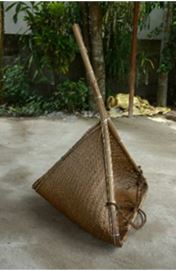
Chiranjit Baruah, from the institute of India. Runu Swargiary, from the institute of India. Hangsha Barman, from the institute of India. Himanga Das, from the institute of India . and Khan Mahammad Khalid, from the institute of India. wrote a Research Article about, Fishing Gears and Their Impact on Aquatic Ecosystems in Barpeta District, Assam. Entitled, Fishing gears, catch composition and their effects on aquatic bodies of Barpeta District, Assam, India. This research paper published by the Journal of Biodiversity and Environmental Sciences (JBES). an open access scholarly research journal on Biodiversity. under the affiliation of the International Network For Natural Sciences | INNSpub. an open access multidisciplinary research journal publisher.

Abstract

Different types of fishing gears used in some localities of Barpeta district, Assam, India were studied. The various fish species caught by the gears and the effects of the gears on habitats of fishes were also studied. It was found that twenty different fishing gears were used by the fishermen of the study area. Among the active gears, there were 3 types of nets, 3 active traps, 3 impaling gears (spears and spikes) and 2 types of hooks and lines. The passive gears included 3 types of gill nets and 2 types of lift nets; 3 types of bamboo traps and 1 barrier. The fishing gears of the study area excluding the surrounding nets and gill nets do not seem to have any detrimental effects on the habitats of fishes or other aquatic organisms.

Submit your article to JBES Journal

Read moreMolecular Characterization of Aspergillus flavus in Imported Maize at Gazetted and UngazettedEntry Points in Kenya | InformativeBD

Introduction

Human food and nutrition is greatly dependent on fisheries and aquaculture, which is also the fastest growing food production industry in the world (Elvarasan, 2018). Inland fishery has a great contribution towards individuals, society and environment. It contributes over 40% of the total world’s finfish production, providing food for millions of people worldwide (Lynch et al., 2016).

Fishing gears, catch composition and their effects on aquatic bodies of Barpeta District, Assam, India

It is seen that freshwater habitats of fish are facing adverse effects of erfishing, destruction and pollution (Yin et al., 2022). In addition, use of destructive fishing gears indiscriminate fishing of juvenile and brood fishes during breeding season also pose a threat to fish diversity (Malakar and Boruah, 2017; Sayeed et al. 2014). Fishing gears like small mesh gill nets and seine nets are destructive to brood fishes, juveniles, fries and fingerlings may lead to loss of fish population. Indiscriminate capture of wild fish populations also has harmful effects on fish biodiversity. The dragging effect of fishing nets also destroys the river bed (Laxmappa and Bakshi, 2014; Kokate et al., 2016; Mia et al., 2017). Reduction of DO due to decomposition of crops due to flood, turbidity, erosion, progressive siltation, destruction of breeding, nursery, grazing fields are some of the natural causes of decline of fish population (Mia et al., 2017). Anthropogenic activities like changing land use practices, increasing population, agricultural expansion and pollution are also increasingly affecting river basins and riverine ecosystems (Mohite and Samant, 2013). It was found that abandoned, lost or discarded fishing gears may contribute significantly to the plastic pollution of Rivers (Nelms et al., 2021).

Fishing gears, catch composition and their effects on aquatic bodies of Barpeta District, Assam, India

Various methods are used by people to catch and aggregate fishes (Sharma et al., 2015). There are many fishing gears which are used in Assam (Pravin et al., 2011; Islam et al., 2013; Baruah, 2014; Purkayastha and Gupta, 2014; Sharma et al., 2017; Jabeen and Soren, 2021; Basumatary and Khangembam, 2023; Borah, et al., 2023). The study was conducted in Barpeta district of Assam, India. Barpeta district has many fishery resources including natural and culture ponds, beels (lakes), rivers and also many swamps and marshy areas. The fishery sector plays a major role in the local economy of the region (Rajbongshi et al., 2016).

Fishing gears, catch composition and their effects on aquatic bodies of Barpeta District, Assam, India

Fishermen use different gears and methods depending on species and environmental and ground conditions. The fishing gears also differ in structure, materials used for construction, capture process and methods of operation. Fishing gears may also have some detrimental effects on the habitats of fishes (Boopendranath, 2009). The present study was done to know the various types of fishing gears used by the fishermen, their local names and the composition of catch and the effects of fishing gears on aquatic water bodies of the study area.

Reference

Baruah D. 2014. Indigenous bamboo-made fishing implements of Assam. Journal of Krishi Vigyan 3(1), 37-41.

Basumatary N, Khangembam BK. 2023. Traditional fishing gears and methods of the Bodo tribes of Kokrajhar, Assam. Fishery Technology 60, 25–36.

Boopendranath MR. 2009. An overview of fishing gears and their design and construction. In: Handbook of Fishing Technology (Meenakumari B, Boopendranath MR, Pravin P, Thomas SN, and Edwin L, Eds). Central Institute of Fisheries Technology, Cochin: 31-66.

Borah GA, Nasreen S, Dutta PK. 2023. A study on ichthyofaunal diversity and fishing gears used in the wetlands areas nearby Nimati Ghat, Jorhat, Assam. Biosciences Biotechnology Research Asia 20(3), 1015-1021.

Elvarasan K. 2018. Importance of fish in human nutrition. Training Manual on Seafood Value Addition, ICAR-Central Institute of Fisheries Technology.

Islam MR, Das B, Baruah D, Biswas SP, Gupta A. 2013. Fish diversity and fishing gears used in the Kulsi River of Assam, India. Annals of Biological Research 4(1), 289-293.

Jabeen F, Soren AD. 2021. Fishing crafts and gears of the river Manas in Assam, India. In: Advances in Scientific Approach for Sustainable Development (Barthakur M, Borthakur MK, Eds.). AkiNik Publications, 172-184.

Kokate AA, Bhosale BP, Metar SY, Chogale ND, Pawar RA, Nirmale VH. 2016. Indigenous fishing crafts and gears of Krishna river with respect to Sangli district of Maharashtra, India. International Journal of Fisheries and Aquatic Studies 4(6), 434-438.

Laxmappa B, Bakshi RR. 2014. Types of fishing gears operating and their impact on Krishna river fishery in Mahabubnagar district, T.S. India. International Journal of Fisheries and Aquatic Studies 2(1), 30-41.

Lynch AJ, Cooke SJ, Deines AM, Bower SD, Bunnell DB, Cowx IG, Nguyen VM, Nohner J, Phouthavong K, Riley B, Rogers MW, Taylor WW, Woelmer W, Youn SJ, Beard TD. 2018. The social, economic, and environmental importance of inland fish and fisheries. Environmental Reviews 24(2), 115-121.

Malakar M, Boruah S. 2017. Diversity and present status of fish species in three floodplain wetlands of Central Assam, India. IOSR Journal of Environmental Science, Toxicology and Food Technology 1(11), 54-59.

Mia M, Islam MS, Begum N, Suravi IN, Ali S. 2017. Fishing gears and their effects on fish diversity of Dekar Haor in Sunamgonj District. Journal of Sylhet Agricultural University 4(1), 111-120.

Mohite SA, Samant JS. 2013. Impact of environmental change on fish and fisheries in Warna River Basin, Western Ghats, India. International Research Journal of Environment Sciences 2(6), 61-70.

Nelms SE, Duncan EM, Patel S, Badola R, Bhola S, Chakma S, Chowdhury GW, Godley BJ, Haque AB, Johnson JA, Khatoon H, Kumar S, Napper IE, Niloy MNH, Akter T, Badola S, Dev A, Rawat S, Santillo D, Sarker S. 2021. Riverine plastic pollution from fisheries: Insights from the Ganges River system. Science of the Total Environment 756, 143305. https://doi.org/10.1016/j.scitotenv.2020.143305.

Pravin P, Meenakumari B, Baiju M, Barman J, Baruah D, Kakati B. 2011. Fish trapping devices and methods in Assam – A review. Indian Journal of Fisheries 58(2), 127-135.

Purkayastha P, Gupta S. 2014. Traditional fishing gears used by the fisherfolk of Chatla floodplain area, Barak Valley, Assam. Indian Journal of Traditional Knowledge 13(1), 181-186.

Rajbongshi MK, Das J, Dutta RK. 2016. Water quality assessment of capture and culture fishery in Barpeta District, Assam, India. International Journal of Fisheries and Aquatic Studies 4(5), 516-520.

Sayeed MA, Hashem S, Salam MA, Hossain MAR, Wahab MA. 2014. Efficiency of fishing gears and their effects on fish biodiversity and production in the Chalan Beel of Bangladesh. European Scientific Journal 10(30), 294-309.

Sharma B, Rout J, Swain SK. 2017. Traditional fishing gadgets used by fishermen of Barak Valley, Southern Assam, North East India. Journal of Entomology and Zoology Studies 5(5), 1555-1560.

Sharma P, Sarma J, Sarma D, Ahmed S, Phukan B, Baishya S, Kashyap D, Dutta MP, Deka P, Hussain IA. 2015. An indigenous fish aggregating method practiced along the Kolong River in Nagaon District of Assam. Indian Journal of Traditional Knowledge 1(1), 112-117.

Yin S, Yi Y, Liu Q, Luo Q, Chen K. 2022. A review on effects of human activities on aquatic organisms in the Yangtze River Basin since the 1950s. River 1, 104-119. https://doi.org/10.1002/rvr2.15.

Source Fishing gears, catch composition and their effects on aquatic bodies of Barpeta District, Assam,India 

Molecular Characterization of Aspergillus flavus in Imported Maize at Gazetted and Ungazetted Entry Points in Kenya | InformativeBD

Molecular characterisation of Aspergillus flavus on imported maize through gazetted and ungazetted points of Entries in Kenya

Joseph Oduor Odongo, from the institute of Kenya. Paul O. Angi’enda, from the institute of Kenya. Bramwel Wanjala, from the institute of Kenya. Catherine Taracha, from the institute of Kenya. and David M. Onyango, from the institute of Kenya. wrote a Research Article about, Molecular Characterization of Aspergillus flavus in Imported Maize at Gazetted and Ungazetted Entry Points in Kenya. Entitled, Molecular characterisation of Aspergillus flavus on imported maize through gazetted and ungazetted points of Entries in Kenya. This research paper published by the International journal of Microbiology and Mycology (IJMM). an open access scholarly research journal on Microbiology. under the affiliation of the International Network For Natural Sciences | INNSpub. an open access multidisciplinary research journal publisher.

Abstract

Maize is a vital staple crop in Kenya, serving as a primary source of food and feed. Contamination of maize (Zea mays) by Aspergillus flavus  and the subsequent production of aflatoxins pose significant threats to food safety and human health. The risk of A. flavus contamination on imported maize at both gazetted and un-gazetted points of entry has not been extensively studied. The primary objective of this study was to examine the genotypic, phenotypic, and aflatoxigenic traits of A. flavus biovars derived from imported maize at Gazetted and Un-gazetted Points of Entries in Kenya. Furthermore, the study sought to establish the phylogenetic relationships among the identified A. flavus strains. A total of 600 imported maize samples were tested for aflatoxin contamination using the Total aflatoxin ELISA test. Out of 600 samples, 4.17% tested positive and were further subjected to morphological and molecular studies.  The morphological analysis revealed the presence of 13 biovars of A. flavus. Micro-morphologically, variations were observed in spore color, size, structure, conidiophore structure, and vesicle shape. The specific primers Calmodulin (CaM), the ITS1-5.8S-ITS2 region of the ribosomal DNA was successfully amplified in 10 out of the 13 biovars that were presumed to be A. flavus, confirming their positive identification as A. flavus. A single band of approximately 700 bp, which corresponds to the expected size of the ITS region in Aspergillus flavus, was observed in 10 out of the 13 biovars. This indicates the presence of A. flavus DNA in those biovars. The amplification of the ITS region provides a specific molecular marker for the identification of A. flavus. These findings highlight the significance of aflQ (ordA) and aflD (nor-1) genes as reliable markers for evaluating the aflatoxigenic potential of A. flavus biovars. Regarding aflatoxigenicity, DV-AM   method was used, and qualitative analysis was conducted. Out of the 13 biovars of A. flavus biovars tested, 23.08% exhibited aflatoxigenicity, while the remaining 10 biovars did not show any aflatoxigenicity. These findings indicate the presence of both aflatoxigenic and non-aflatoxigenic strains of A. flavus among the imported maize samples. The phylogenetic analysis revealed that Taxon 31 (AY495945.1 Aspergillus flavus biovar 92016f aflR-aflJ intergenic region partial sequence) and Taxon 32 (NR 111041.1 Aspergillus flavus ATCC 16883 ITS region from TYPE material). This genotypic and phenotypic characterization provides valuable information for understanding the diversity and potential toxigenicity of A. flavus strains on imported maize. This study contributes to the understanding of the genotypic and phenotypic characteristics of A. flavus on imported maize in Kenya.

Submit your article to IJMM Journal

Read moreIsolation and Identification of Bacterial Pathogens in Infected Shing Fish (Heteropneustesfossilis) from Freshwater Ponds in Bangladesh | InformativeBD

Introduction

Maize plays a central role in the food security and livelihoods of Kenyan populations. It serves as a staple food crop for a significant portion of the population, contributing to both dietary needs and income generation. Moreover, maize is an essential component of livestock feed, supporting the growth of the domestic livestock industry. In sub-Saharan Africa as a whole, maize is ranked third in importance among cereal crops, following rice and wheat (Shiferaw et al., 2011). The cultivation and trade of maize have a considerable impact on regional economies and food systems. Maize (Zea mays) is often contaminated by Aspergillus fungal species during pre- and post-harvest practices, storage, and transportation. Studies by Horn (2007) showed that Aspergillus species are commonly found in the soil, which acts as a source of primary inoculum for infecting developing maize kernels during the growing season. Aspergillus flavus is distributed globally with a high frequency of occurrence in warm climates which favor the growth of the fungus (Cotty et al., 1994).

Understanding the population structure and genetic diversity of A. flavus is crucial for diversification of effective management strategies. Different strains of A. flavus may have varying levels of aflatoxin production and pathogenicity, which can influence the severity of contamination in maize (Abbas et al., 2013). Additionally, certain strains may exhibit resistance or susceptibility to control measures, such as biological control agents or fungicides. Therefore, identifying specific strains or groups within the A. flavus population can aid in the selection of appropriate control strategies to minimize aflatoxin contamination. Moreover, the genetic diversity of A. flavus may also have implications for host-pathogen interactions and disease development. Different strains may exhibit variations in their ability to infect maize kernels, colonize host tissues, and compete with other microorganisms in the maize ecosystem (Atehnkeng et al., 2014). Understanding these interactions can help in the development of resistant maize varieties and cultural practices that can limit fungal growth and subsequent aflatoxin production. The population structure and genetic diversity of A. flavus strains isolated from maize play a significant role in aflatoxin contamination and disease development. The existence of multiple strains within the A. flavus population highlights the need for comprehensive investigations to characterize their phenotypic and genotypic traits. Such studies will provide insights into the factors influencing aflatoxin production, the design of effective control strategies, and the development of resistant maize varieties to minimize the health and economic risks associated with aflatoxin contamination. Aspergillus species, including Aspergillus flavus, are of great concern due to their ability to produce aflatoxins, potent carcinogens and toxins that contaminate various agricultural commodities, including maize. The accurate identification and characterization of Aspergillus species is crucial for assessing their potential to produce aflatoxins and understanding their impact on food safety.

Gene sequencing has emerged as a powerful tool for the accurate identification and classification of Aspergillus species. In recent years, numerous studies have utilized gene sequencing data to characterize Aspergillus biovars from different sources. By comparing the genetic sequences of specific genes, such as the internal transcribed spacer (ITS) region, researchers can determine the species and genetic diversity within a population. In addition to genetic characterization, a polyphasic approach is commonly employed to identify and characterize Aspergillus biovars. This approach combines morphological and molecular analyses to provide a comprehensive understanding of the biovars. Morphological characteristics, such as colony color, texture, spore color, size and structure, conidiophore structure and vesicle shape are observed and recorded. These characteristics help in differentiating between various Aspergillus species and subgroups. Furthermore, molecular techniques, including polymerase chain reaction (PCR) amplification and sequencing of specific genetic markers, allow for a more precise identification of aflatoxigenic and nonaflatoxigenic A. flavus biovars. These methods target genes associated with aflatoxin production, such as the aflatoxin biosynthesis cluster genes, to determine the potential of a biovar to produce aflatoxins. The combination of gene sequencing and polyphasic approaches provides a comprehensive understanding of the genetic diversity, population structure, and aflatoxinproducing potential of Aspergillus species, particularly A. flavus. This information is essential for risk assessment, development of effective control strategies, and ensuring the safety and quality of imported maize and other agricultural commodities.

This study contributed to the understanding of the population dynamics and potential risks associated with A. flavus in imported maize. Given the prominence of maize in Kenya, research efforts focusing on this crop are crucial. The genotypic and phenotypic characterization of A. flavus on imported maize assumes particular significance in the Kenyan context. A thorough understanding of the genetic diversity and potential for mycotoxin production in A. flavus populations is essential for developing effective control strategies and mitigating the health risks associated with mycotoxin contamination. Gazetted and un-gazetted points of entry play a crucial role in facilitating the importation of maize. However, the risk of A. flavus contamination in imported maize has not been thoroughly investigated, warranting a comprehensive genotypic and phenotypic characterization of this fungus. Understanding the genotypic and phenotypic characteristics of A. flavus on imported maize is essential for several reasons. Firstly, it allows for the identification of specific genetic traits and phenotypic features associated with higher aflatoxin production, thus enabling the development of targeted control strategies. Secondly, it provides insights into the diversity of A. flavus biovars present in imported maize and their potential for aflatoxin contamination. This knowledge can contribute to risk assessment and management strategies aimed at preventing or minimizing aflatoxin contamination in the domestic maize supply chain.

Genotypic characterization involves studying the genetic makeup of A. flavus biovars to determine their relatedness, genetic diversity, and potential for toxin production. Several molecular techniques have been used for genotyping A. flavus, including random amplified polymorphic DNA (RAPD), amplified fragment length polymorphism (AFLP), and multilocus sequence typing (MLST) (Abdallah et al., 2018). These methods have provided valuable insights into the genetic diversity and population structure of A. flavus, highlighting the presence of distinct genotypes in different geographic regions (Klich et al., 2015). Phenotypic characterization involves studying the observable traits and behaviors of A. flavus, such as growth patterns, conidiation, and mycotoxin production. Phenotypic characterization is essential for understanding the pathogenicity and virulence of A. flavus strains on imported maize. Researchers have observed variations in colony morphology, growth rate, and sporulation among different A. flavus biovars (Calvo et al., 2016). Furthermore, studies have demonstrated the production of mycotoxins, particularly aflatoxins, by certain A. flavus strains (Chang et al., 2019). Phenotypic characterization provides valuable information for risk assessment and identifying high-risk A. flavus biovars in imported maize. The genotypic and phenotypic characterization of A. flavus on imported maize plays a crucial role in assessing the potential health risks associated with mycotoxin contamination. By combining genotypic and phenotypic data, researchers can identify highly toxigenic A. flavus strains and evaluate their prevalence in imported maize.

This information is essential for implementing targeted control measures, such as crop management strategies, post-harvest interventions, and storage practices, to minimize mycotoxin contamination and ensure food safety (Li et al., 2020). Investigating A. flavus on imported maize specifically at gazetted and ungazetted points of entry in Kenya is crucial. Gazetted points of entry are official border checkpoints designated for the importation of agricultural products, while un-gazetted points of entry refer to informal channels through which goods, including maize, are smuggled into the country. Analyzing both types of entry points can provide a comprehensive understanding of the risks associated with A. flavus contamination in imported maize, as well as the efficacy of control measures implemented at official checkpoints. In this study, we aim to conduct a detailed genotypic and phenotypic characterization of A. flavus on imported maize at both gazetted and un-gazetted points of entry in Kenya. We will analyze the genetic diversity, aflatoxin production capability, and other phenotypic traits of A. flavus biovars obtained from imported maize samples. By doing so, we hope to gain insights into the potential sources and pathways of A. flavus contamination in imported maize and develop targeted strategies to ensure the safety and quality of imported maize in Kenya.

Reference

Abbas HK, Accinelli C, Zablotowicz RM, Abel CA, Bruns HA. 2013. Prevalence of aflatoxin and fumonisin in corn (maize) and peanut cake from hens laying contaminated eggs destined for human consumption in Pakistan. Food Additives & Contaminants: Part A, 30(1), 169-180. DOI:10.1080/19440049.2012.748706

Abdallah MF, Girgis GN, Khedr AHA, Ali EF, Abdul-Raouf UM. 2018. Genotypic diversity and antifungal susceptibility of Aspergillus flavus isolated from maize grains in Egypt. Journal of Genetic Engineering and Biotechnology, 16.

Amaike S, Keller NP. 2011. Aspergillus flavus. Annual Review of Phytopathology 49, 107-133.

Atehnkeng J, Donner M, Ojiambo PS, Ikotun T, Sikora RA, Cotty PJ, Bandyopadhyay R. 2014. Biological control agents for managing aflatoxin contamination in groundnut: A review. Agriculture 4(3), 197-217. DOI: 10.3390/agriculture4030197

Atehnkeng J, Ojiambo PS, Ikotun T, Sikora RA, Cotty PJ, Bandyopadhyay R. 2014. Evaluation of atoxigenic isolates of Aspergillus flavus as potential biocontrol agents for aflatoxin in maize. Food Additives & Contaminants: Part A, 31(2), 378-387.

Bensch K, Groenewald JZ, Dijksterhuis J, Starink-Willemse M, Andersen B, Summerell BA, Shin HD, Dugan FM. 2018. Species and ecological diversity within the Cladosporium cladosporioides complex (Davidiellaceae, Capnodiales). Studies in Mycology 89, 177-301.

Brown DW. 2005. Phylogenetic Analysis of Zipper-Positive Abdominal Aflatoxin-Producing Aspergillus Species. Mycologia 97(2), 498-504.

Brown DW. 2010. Phylogenetic Analysis and Mycotoxin Production Capability of Aspergillus Section Flavi from Brazil Nuts. Mycologia 102(4), 866-872.

Chen ZY, Brown RL, Rajasekaran K, Damann KE, Cleveland TE. 2008. Evaluation of thermotolerant strains of Aspergillus flavus for aflatoxin contamination and genetic variation. Journal of Food Protection 71(9), 1909-1914.

Diba K, Kordbacheh P, Mirhendi SH, Rezaie S, Mahmoudi M. 2007. Identification of Aspergillus species using morphological characteristics. Pakistan Journal of Medical Sciences 23(6), 867.

Diniz LE, Sakiyama NS, Lashermes P, Caixeta ET, Oliveira ACB, Zambolim EM, Zambolim L. 2005. Analysis      of AFLP markers associated to the Mex-1 resistance locus in Icatu progenies. Crop Breeding And Applied Technology, 5(4), 387.

Johnson LJ. 2015. Genetic Diversity and Population Structure of Aspergillus flavus Isolates from Maize Fields in Three Geographic Regions of the United States. Journal of Agricultural and Food Chemistry 63(41), 9016-9023.

Jones JP. 2012. Molecular identification of aflatoxigenic and non-aflatoxigenic Aspergillus species from maize (Zea mays L.). Mycotoxin Research 28(2), 89-96.

Kilonzo RM, Imungi JK, Muiru WM, Lamuka PO, Kuria EN. 2017. Genetic diversity and aflatoxin contamination of maize from eastern Kenya regions. Journal of Applied Biosciences 114, 11342-11351.

Kilonzo-Nthenge A, Monda E, Okoth S, Makori D. 2019. Aflatoxins and their fate in maize and maize-based products in Kenya: A review. Food Control 96, 219-225.

Kilonzo-Nthenge A, Monda E, Okoth S, Makori D. 2019. Aflatoxins and their fate in maize and maize-based products in Kenya: A review. Food Control 96, 219-225.

Lee T. 2019. Genetic Diversity and Population Structure of Aspergillus flavus Isolates from Maize in Thailand. Frontiers in Microbiology 10, 1997.

Liang Y, Yu J, Zhou T. 2015. Improving Aflatoxin B1 Production on Rice by Aspergillus flavus and Aspergillus parasiticus through Recombination of Cytochrome P450 Enzymes. Scientific Reports 5, 1–10. https://doi.org/10.1038/srep08260

Miller JD. 2016. Structure and Population Diversity of Aspergillus flavus from Maize in Thailand. Toxins 8(12), 368.

Odhiambo BO, Kilonzo RM, Njage PM, Okoth S. 2020. Aflatoxin contamination in maize: Current challenges and potential opportunities for mitigation in Sub-Saharan Africa. Toxins 12(10), 630.

Ojiambo PS, Ikotun T, Leke W, Sikora R, Mukalazi J. 2016. Variation in the pathogenic and genetic diversity among isolates of Aspergillus flavus link to aflatoxin contamination of peanut and maize in Kenya.

Probst C, Schulthess F, Cotty PJ. 2010. Impact of Aspergillus section Flavi community structure on the development of lethal levels of aflatoxins in Kenyan maize (Zea mays). Journal of Applied Microbiology 108(2), 600-610. DOI: 10.1111/j.1365-2672.2009.04432.x

Raju MVLN, Seetharami Reddi TV, Krishna TG. 2014. Development of a PCR-based method for detection and differentiation of Aspergillus flavus and Aspergillus parasiticus. Indian Journal of Microbiology 54(2), 202-206.

Samson RA, Houbraken J, Thrane U, Frisvad JC, Andersen B. 2019. Food and indoor fungi. Westerdijk Fungal Biodiversity Institute.

Samson RA, Houbraken J, Thrane U, Frisvad JC, Andersen B. 2014. Food and Indoor Fungi. CBS-KNAW Fungal Biodiversity Centre.

Sobolev VS, Neff SA, Gloer JB, Abbas HK. 2009. Characterization of novel volatile antimicrobials from the molds Aspergillus flavus, Aspergillus parasticus, and Aspergillus ochraceus. Toxins 1(1), 3-12. DOI: 10.3390/toxins1010003

White TJ. 1990. Amplification and Direct Sequencing of Fungal Ribosomal RNA Genes for Phylogenetics. In PCR Protocols: A Guide to Methods and Applications (pp. 315-322). Academic Press.

Xu J, Chen AJ, Zhang Y. 2016. Molecular approaches for rapid identification and diversity assessment of foodborne spoilage yeasts: A review. Journal of Food Science 81(1), M15-M22.

Yu J. 2016. Regulation of Aflatoxin Biosynthesis: Perspectives from Genomics Research. Methods in Molecular Biology 1398, 267-285.

Yu J, Chang PK, Cary JW, Wright M, Bhatnagar D, Cleveland TE, Payne GA. 2012. Comparative mapping of aflatoxin pathway gene clusters in Aspergillus parasiticus and Aspergillus flavus. Applied and Environmental Microbiology, 78(23), 7856-7866. DOI: 10.1128/AEM.01959-12

Yu J, Chang PK, Ehrlich KC, Cary JW, Bhatnagar D, Cleveland TE, Payne GA. 2004. Clustered pathway genes in aflatoxin biosynthesis. Applied and Environmental Microbiology 70(3), 1253-1262.

Yu J, Chang P-K, Ehrlich KC, Cary JW, Montalbano B, Dyer JM, Bhatnagar D, Cleveland TE, Payne GA. 2011. Clustered pathway genes in aflatoxin biosynthesis. Applied and Environmental Microbiology 77(24), 8479-8484. https://doi.org/10.1128/AEM.06367-11

Yu J, Payne GA, Nierman WC, Machida M, Bennett JW, Campbell BC. 2013. Aspergillus flavus genomics: Gateway to human and animal health, food safety, and crop resistance to diseases. In Advances in Applied Microbiology  84, 1-91. 

Source Molecular characterisation of Aspergillus flavus on imported maize through gazetted and ungazetted points of Entries in Kenya