Plant-Parasitic Nematodes in Peanut Fields | InformativeBD

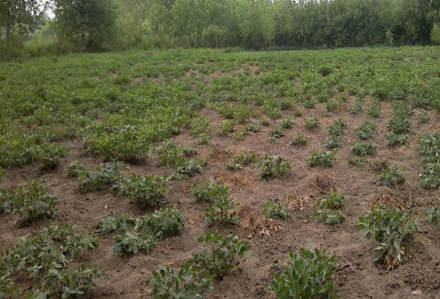
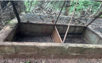
Field symptoms showing patchy distribution of damage caused by groundnut plants infected byHeterodera cruciferea.

Seyede Negin Mirghasemi, Mina neginfar, Salar Jamali , Mina Allamoradi  and Amaneh Hosseinikhah Choshali from the different institute of the Iran, wrote a research article about, Plant-Parasitic Nematodes in Peanut Fields, entitled,  "Reported some species of plant parasitic nematodes from rhizosphere of peanut (Arachis ypogaea) fields". This research paper published by the International Journal of Mycrobiology and Mycology | IJMM. an open access scholarly research journal on Mycrobiology, under the affiliation of the International Network  For Natural Sciences | INNSpub. an  open access multidisciplinary research journal publisher.

Abstract

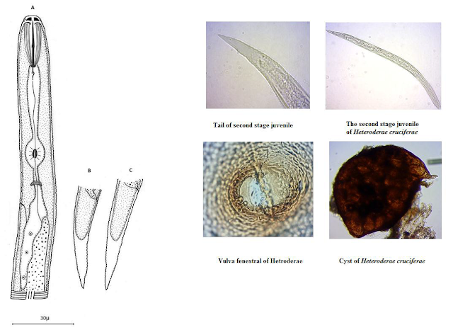
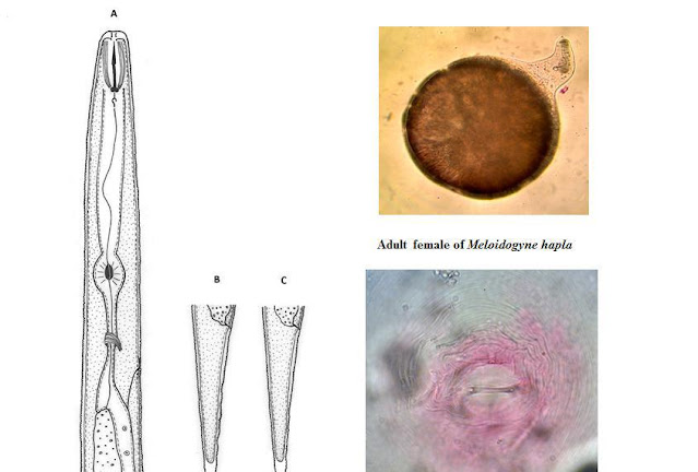
In order to identify of peanut fields plants parasitic nematodes, 130 samples of soil around the roots of peanut plants were collected in province of Guilan, during the summer and fall of 2011. After extraction, killing, fixation and transferring to anhydrous glycerol, the nematodes were mounted on permanent microscopic slides and nematodes species identified by using light microscope, equipped with digital camera, based on morphological and morphometric characters using valid keys. In this study 20 species belonging 17 genera were identified, that are as followes: 1- Aphelenchoides sacchari 2-Aphelenchus avenae 3- Basiria graminophila 4-Coslenchus costatus 5-Ditylenchus myceliophagus 6-Filenchus vulgaris 7-Helicotylenchus digonichus 8-Heterodera cruciferae 9-Meloidogyne incognita 10-Meloidogyne hapla 11-Merlinius bavaricus 12- Mesocriconemarusticum13- Mesocriconema curvatum 14-Paratylenchus nanus 15-Pratylenchus neglectus 16- Psilenchus hilarulus 17-Quinsulcius capitatus 18-Tylenchorhynchus annulatus 19- Tylenchorhynchus mashhoodi 20- Tylenchus davainei. In thisstudy, 20 species belonging 17 genera were identified that before just 6 Species2-5-7-11-15-16were reported from rhizosphere of Peanut in Iran. Other species (14 Species) are going to report from rhizosphere of Peanut in Iran as a first.

Submit your article to IJMM Journal

Read more Caladium bicolor: In Vitro Regeneration Insights | InformativeBD

Introduction

Arachis hypogaea, the peanut or groundnut, is an annual herbaceous plant in the Fabaceae (legume or bean family), with high protein content, vitamins and useful mineral compounds. Total commercial production of peanuts worldwide was 37.6 million tons, harvested from 24.1 million hectares, in 2010. Iran cultivated area The most under cultivating area is located in Guilan province of Iran as 90 present of peanut crop were cultivated in this area (Anon,2012).

Reported some species of plant parasitic nematodes from rhizosphere of peanut (Arachis ypogaea) fields

Peanut Arachis hypogaea is considered one of the important food crops, produced in many subtropical and tropical countries; also it is a high value cash crop for small and large growers alike. It was listed as one of the twenty crop plants that stand between man and starvation (Wittwer, 1981). Numerous nematode species can damage peanut.(Minton &Baujard, 1990).Several plant parasitic nematodes were reported from peanut fields in South Eastern States of USA such as Belolaimus longicaudatus, Meloidogyne hapla, Meloidogyne arenaria, Mesocriconema ornatum and Pratylenchus brachyurus (Barker, 1992). Peanut root–knot nematode Meloidogyne arenaria is considered one of the most serious nematode pathogens of peanut in many parts of the world, attacking peanut roots, pegs and pods and causing substantial yield losses in severely infested fields (Minton & Baujard, 1990). Meloidogyne arenaria and M. hapla are the most important peanut crop loss agent in USA, which their populations have reached as economically damaging threshold (Hirunsalee et al.,1995). Elekcioglu et al. (1994) also have reported many species Aphelenchus avenae, Ditylenchus myceliophagus, D. valveus, Pratylenchus thornei, Tylenchorhynchus goffartri from peanut fields in the east mediterranean region of Turkey.

Reported some species of plant parasitic nematodes from rhizosphere of peanut (Arachis ypogaea) fields

In order to improve cultivating steps in peanut and prevent the decline of peanut crop production in traditional cultures, the need to identify plant pathogens, is the first and most important step in disease management. Since the yield loss associated with plant pathogens including plant parasitic nematodes, this study aims to investigate the peanut nematodes.

Reference

Barker KR. 1992. Effects of Meloidogyne hapla and Meloidogyne arenaria on black rot severity in new Cylindrodadium-resistant peanut genotypes. Plant Disease, 76, 352-357.

Brzeski WM. 1998. Nematodes of Tylenchina in Poland and temperate Europe. Muzem I Inst. Zool. PolskaAkad.Nauk.Warsaw, Poland, 396 P.

Brzeski MW, Dolinski CM. 1998. Compendium of the genus Tylenchorhynchus Cobb, 1913 sensulato (Nematoda: Belonolaimidae). Russian Journal of Nematology6:189-199. (Synonymy of six genera with Tylenchorhynchus).

Coolen WA, D´herde CJ. 1972. A method for the quantitative exttissue. Ghent: State Nematology and Entomology Research Station, 77p.raction of nematodes from plant.

De Grisse A. 1969. Redescription ou modification de quelques dans letude des nematodes Phytoparasit. Mededelingen Rijksfaculteit der landbouwwetenschappen Gent, 34, 351-369.

Elekcioglu˙IH, Osnesorge B, Lung G, Uygun N. 1994. Plant parasitic nematodes in the East Mediterranean Region of Turkey. Nematol. Medit. 22, 59-63.

Geraert E. 2010. The Criconematidae of the world, Identification of the family Criconematidae (Nematoda). Academia Press, Gent, Belgium.pp. 615.

Handoo ZA. 2000. A Key and Diagnostic Compendium to the Species of the Genus Tylenchorhynchus Cobb, 1913(Nematoda: Belonolaimidae). Journal of Nematology, 32(1), 20-34.

Handoo Z, Skantar AM, Carta LK, Schmitt DP. 2005. Morphological and Molecular Evaluation of MeloidogynehaplaPopulation Damaging Coffee (Coffee arabica) in Maui, Hawaii.Journal of Nematology, 37(2), 136-145.

Hirunsalee A, Barker KR, Beute MK. 1995. Infection, Reproduction Potential, and Root Galling by Root-Knot Nematode Species and Concomitant Population on Peanut and Tobacco.Jurnal of Nematology, 27(2), 172-177.

Jepson S. 1987.Identification of root-knot nematodes (Meloidogynespecies). London, UK. C.A.B International, 293p.

Minton NA, Bajuard P. 1990. Nematode parasite of peanut.Pp: 285-320. In: Plant Parasitic Nematodes in Subtropical and Tropical Aagriculture (M. Luc, R.A. Sikora& J. Bridge, eds). CAB.International Inst. Parasitology.Wallingford Oxon. UK.

Raski DJ. 1957. Revision of the Genus Paraty lenchus Micoletzky, 1922, and describtions of new species. Part 2 of Three parts. J. Nematol.7, 274-295.

Wittwer SH. 1981. The 20 corps that stand between man and starvation. Farm chemicals, 144, 17-28.

Wouts WM. 2006. Fauna New Zealand Kote Atitanaga Pepeke o Aotearoa Criconematina (Nematoda: Tylenchida).Lincoln, Canterbury, New Zealand, 55, 232pp.

anonymous.2011.(http://www.iran.ir/about/city /gilan. 5/12/2012).

anonymous.2009.Ebtekarnews.(http://www.ebt ekarnews.com/ebtekar/news.aspx?nid=58380.5/ 12/2012).

Anonymous. 2000-2012. Soya tech, growing opportunities. (http://www.soyatech.com/peanut_facts.htm.5/1

2/2012).

 Source: Reported some species of plant parasitic nematodes from rhizosphere of peanut (Arachis ypogaea) fields


Caladium bicolor: In Vitro Regeneration Insights | InformativeBD

 Maximum multiple shoot induction at 0.5mg/L BA + 2.0mg/L IBA supplemented MS media

KS. Ahmed,  ME. Hoque , M. Shamsuzzaman, S. Sultana, and MS. Islam from the different institute of the dhaka, wrote a research article about, Caladium bicolor: In Vitro Regeneration Insights, entitled,"In vitro regeneration of Caladium bicolor".This research paper published by the International Journal of Biosciences | IJB. an open access scholarly research journal on Biosciences. under the affiliation of the International Network For Natural Sciences | INNSpub. an open access multidisciplinary research journal publisher.

Abstract

The present experiment was conducted to determine the ideal concentration of different plant growth regulators (BA, Kin, IBA, IAA, and NAA) for in vitro regeneration of Caladium bicolor using shoot tip explants. The work was designed in CRD with three replications. Shoot tip explants gave rise to multiple shoots when cultured on MS medium supplemented with different concentration of BA with IBA. The highest (90%) response of shoot multiplication was obtained in MS medium containing 0.25-1.0mg/L BA + 2.0-2.5mg/L IBA. The regenerated shoots were then rooted on MS medium with different concentrations NAA, IAA and IBA. The maximum frequency of rooting and highest number of roots was produced on medium containing 2.0mg/L IAA. In accordance with average growth characteristics, it was revealed that the combined effect of BA and IBA appeared to be better to individual performance. The plantlets, thus developed were hardened and successfully established in soil. The plants raised through tissue culture exhibited normal growth. Reliable protocols for micropropagation of Caladium bicolor were established, which could be used for large scale production of disease free, high-yielding, and premium quality planting material.

Submit your article to IJB Journal

Introduction

Caladium bicolor is a member of Araceae family(arum family) and commonly known as angel wings,heart of Jesus and fancy-leaved caladium (Ali et al.,2007; Syedi et al., 2016). It is an importantornamental plant valued for its long-lasting colorfulfoliage, and is commonly grown in containers and inthe landscape (Syedi et al., 2016; Deng, 2018; Zhanget al, 2019). They are grown for their colorful leavesthat have a combination of green and white, greenand red, white with red blotches or green veins andsome have lavender spots. The size of the heartshapedleaves may vary from 6 inches to 2 feet inlength. Ornamental value of Caladiums depends to agreat extent on leaf characteristics, including shape,color, color pattern, and venation pattern (Deng andHarbaugh, 2005).

In vitro regeneration of Caladium bicolor

Generally, Caladium is propagated from tubers forcommercial purpose but tuber propagation haslimitations as tubers produce healthier plants for oneseason only and second year foliage is usually not asgood as the first year. Therefore, more satisfactoryresults may be obtained by starting with new tuberseach year. Commercial propagation can also beachieved through seeds but the seed propagation isdifficult, being seeds very small, requires handpollinations, very high mortality and very difficult tokeep plant true to type and pathogen free. Moreover,plants grown from seeds are very expensive. It hasalso been reported that seed propagation results invariability (Ali et al., 2007; Deng et al., 2007).Concerns have been raised about possible loss ofgenetic diversity due to a drastic decline in thenumber of cultivars in the last century. Moreover, thismethod is very difficult to keep plant true to type andpathogen free (Siddiqui et al., 1993; Deng et al.,2007). Consequently, seed propagation is not used incommercial production of caladium plants.

In vitro regeneration of Caladium bicolor

Recently, many caladium companies and nurserieshave started using tissue culture technology known asmicropropagation for large scale production of true totype and disease free caladium. In vitro techniquesare powerful tools for plant breeders in improving theperformance of agriculture, horticulture andfloriculture plant species. Interest in tissue culturepropagation of Caladium bicolor has evolved due to itsornamental importance throughout the world. Thesuccess of the micropropagation method depends onseveral factors like genotype, media, PGRs and type ofexplants (Pati et al., 2005; Nhut et al., 2010). Someinvestigations were done on micropropagation ofCaladium spp. using leaf, apical meristem,inflorescences and other explants and a high number oftreatments, plant growth regulators (PGRs), and dosages( Mujib et al., 2000; Chu and Yazawa, 2001; Ahmad etal., 2004; Ali et al., 2007; Thepsithar et al., 2010).

Therefore, the present investigation was carried out to identify the best hormonal combination in Caladium bicolor regeneration as well as rapid and easy in vitro propagation of Caladium bicolor.

Reference

Ahmad EU, Hayashi T, Yazawa S. 2004. Auxins increase the occurrence of leaf-colour variants in Caladium regenerated from leaf explants. Scientia horticulturae 100, 153-9. https://doi.org/10.1016/ j.scienta.2003.08.012

Ali AA, Munawar AS, Naz SH. 2007. An in vitro study on micropropagation of Caladium bicolor. International Journal of Agriculture and Biology 9(5), 731-735.

Al-Taleb MM, Hassawi DS, Abu-Romman SM. 2011. Production of virus free potato plants using meristem culture from cultivars grown under Jordanian environment. American-Eurasian Journal of Agricultural & Environmental Sciences 11(4), 467-72.

Barakat AA, Gaber MK. 2018. Micropropagation and ex vitro acclimatization of aglaonema plants. Sciences 8(4), 1425-36.

Chan LK, Tancm, Chew GS. 2001. Micropropagation of the Araceae ornamental plants. In International Symposium on Acclimatization and Establishment of Micropropagated Plants 616, 383-390.

Chu Y, Yazawa S. 2001. The variation and the hereditary stability on leaf character of plantlets regenerated from micropropagation in Caladiums. Journal of Chinese Society for Horticultural Science 47, 59-67.

Deng Z, Goktepe F, Harbaugh BK, Hu J. 2007. Assessment of genetic diversity and relationships among caladium cultivars and species using molecular markers. Journal of the American Society for Horticultural Science 132(2), 219-29. https:// doi.org/10.21273/JASHS.132.2.219

Deng Z, Harbaugh BK. 2005. Inheritance of leaf shapes and main vein colour in Caladium. U.S. Department of Agriculture, Cooperative Extension Service, University of Florida, IFAS, Florida A. & M; Publication ENH 1006.

Deng Z. 2018. Caladium. In: Van Huylenbroeck, J. (Eds) Ornamental Crops. Handbook of Plant Breeding 11, 273-299. https://doi.org/10.1007/978-3-319-90698-0-12

Dhital SP, Lim HT, Manandhar HK. 2011. Direct and efficient plant regeneration from different explants sources of potato cultivars as influenced by plant growth regulators. Nepal Journal of Science and Technology 12, 1-6. https://doi.org/10.3126 /njst.

Ghasemi Ghehsareh M, Ghanbari M, Reezi S. 2020. The effects of different potted mixtures on the growth and development of miniature roses (Rosa ‘Orange Meillandina). International Journal of Recycling Organic Waste in Agriculture 9(4), 399-409. https://doi.org/10.30486/ijrowa.2020.1897723.1060

Jain SM, Ochatt S. 2010. Protocols for in vitro propagation of ornamental plants. Springer Protocols: Humana press.

Kaviani B, Hashemabadi D, Khodabakhsh H, Onsinejad R, Ansari MH, Haghighat N. 2015. Micropropagation of Begonia rex Putz. by 6-benzyladenine (BA) and α-naphthalene acetic acid (NAA). International Journal of Biosciences 6(5), 8-15. http://dx.doi.org/10.12692/ijb/6.5.8-15

Kaviani B. 2015. Some useful information about micropropagation. Journal of Ornamental Plants 5(1), 29-40.

Mujib A, Bandhyopadhyay S, Ghosh PD. 2000. Tissue culture derived plantlet variation in Caladium an important ornamental. Plant Cell, Tissue and Organ Culture 10, 149-155.

Mujib A, Banerjee S, Fatima S, Ghosh PD. 2008. Regenerated plant populations from rhizome-calli showed morphological and chromosomal changes in Caladium bicolor (Ait.) Vent. cv. Bleeding Heart. Propagation Ornament Plants 8(3), 138-43.

Murashige T, Skoog, FA. 1962. Revised medium for rapid growth and bioassays with tobacco tissue cultures. Physiologia Plantarum 15, 473-497.

Nhut DT, Hai NT, Phan MX. 2010. A highly efficient protocol for micropropagation of Begonia tuberous. In: Jain SM, Ochatt SJ(eds) Protocolsfor In vitro Propagation of Ornamental Plants, Springer protocols. Humana Press. pp. 15-20.

Pati PK, Rath SP, Sharma M, Sood A, Ahuja PS. 2006. In vitro propagation of rose: A review. Biotechnology advances 24(1), 94-114. https://doi.org/10.1016/j.biotechadv.2005.07.001

Pierik RLM. 1987. In vitro culture of higher plants. Dordrecht, The Netherlands, Martinus Nijhoff 45-82.

Sanavy SA, Moeini MJ. 2003. Effects of different hormone combinations and planting beds on growth of single nodes and plantlets resulted from potato meristem culture. Plant Tissue Culture 13(2), 145-50.

Seydi S, Negahdar N, Taghizadeh AR, Ansari MH, Kaviani B. 2016. Effect of BAP and NAA on micropropagation of Caladium bicolor (Aiton) vent., an ornamental plant. Journal of Ornamental Plants 6(1), 59-66.

Siddiqui FA, Naz S, Iqbal J. 1993. In vitro propagation of Carnation. Advances in plant tissue culture. Proceedings of the 3rd National Meeting of Plant Tissue Culture Pakistan pp. 43-7.

Thepsithar C, Thongpukdee A, Chiensil P. 2010. Micropropagation of Caladium bicolor (Ait.) Vent.Thep Songsil’and incidence of somaclonal variants. Acta Horticulturae (855), 273-280.

Van Staden J, Zazimalova E, George EF. 2008. Plant growth regulators II: Cytokinins, their analogues and antagonists. Plant propagation by Tissue culture 1, 205-226.

Webb KJ, Osifo EO, Henshaw GG. 1983. Shoot regeneration from leaflet discs of six cultivars of potato (Solanum tuberosum subsp. tuberosum). Plant Science Letters 30(1), 1-8. https://doi.org/10.1016/0304-4211(83)90196-7

Zhang YS, Gu SJ, Chen JJ, Cai XD. 2019. Effects of different nutrient solutions on the acclimatization of in vitro Caladium plantlets using a simplified hydroponic system. Sains Malays 1(48), 1627-33. http://dx.doi.org/10.17576/jsm-2019-4808-08

 SourceIn vitro regenerationof Caladium bicolor


Optimizing Growth: Diet & Substrate for Archachatina Marginata | InformativeBD


Breeding pen.

Kouassi Kouadio Daniel , N’guessan N’guessan Olivier, and  Aman Jean-Baptiste from the different institute of the Côte d'Ivoire, wrote a research article about Optimizing Growth: Diet & Substrate for Archachatina Marginata, entitled, "Interaction on the diet and substrate on the growth of Archachatina marginata in breeding".This research paper published by the Journal of Biodiversity and Environmental Sciences | JBES. an open access scholarly research journal on Biodiversity. under the affiliation of the International Network For Natural Sciences | INNSpub. an open access multidisciplinary research journal publisher.

Abstract

Nine hundred juveniles of Archachatina marginata aged about two weeks, with an average live weight of 2.25 g with an average shell length of 20.12mm were monitored in culture for six (6) months on five types of substrates [S1 (soil collected in a cassava plantation: Manihot sp.), S2 (S1 with 10% oyster shell meal), S3 (S1 with 10% sawdust), S4 (S1 with 5% oyster shell meal and 5% sawdust) and S5 (uncultivated forest soil). Four diets including two industrial (D1 and D 2 of 12% and 16% calcium respectively) and two based on fodder (D3 and D4 based on leaves and fruit of the papaya (Carica papaya) on the one hand and a mixture of papaya leaves and taro (Xanthosoma maffafa) on the other hand, were used. In order to determine the best combinations inducing the best growth performance, 20 combinations were formed at the rate of 45 spat for each combination; three replicas of 15 spat each. This study showed that the combination of diet and livestock substrate influences the growth of Archachatina marginata. Although the best feed is D1 (74.68 g and 7.94cm) and the best substrate is S2 (77.12 g and 7.79cm), the best combinations are D2S3 (69.37 g and 7.47cm), D1S4 (74.68 g and 7.94cm and D4S2 (77.12 g and 7.79cm). The combined effect of the high level of dietary calcium and that of the culture substrate does not promote good growth of snails. This work will help improve the production of African giant snails and provide important data for anyone wishing to engage in the breeding of these animals. 

Submit your article to JBES Journal

Read more : Blue Butterfly Pea: Phytochemicals and E. coli Inhibition | InformativeBD

Introdction 

Naturally available food resources play a fairly substantial role in populations (Sodjinou et al., 2002). Among these resources, African giant snails (or Achatines) belonging to the family Achatinidae are found there. These snails are highly valued by many African populations (Zongo 1995). For example, Achatine meat is the most consumed meat in South Benin ahead of aulacode, chicken, sheep or goats, beef and pork (Sodjinou et al., 2002). It is estimated that in Côte d'Ivoire, the population eats 7.9 million kg of snails per year, while in Ghana; demand clearly exceeds production capacity (Cobbinah et al., 2008).

Interaction on the diet and substrate on the growth of Archachatina marginata in breeding

Unfortunately, these protein resources are becoming scarce in their natural environment. To compensate for these deficits, heliculture is one of the alternatives to diversify the sources of animal protein of populations. It is therefore right that initiatives to breed these animals should be carried out in order to satisfy the ever-increasing demand for their consumption, but also to ensure the sustainability of the resource. Thus, several research initiatives on the pace of activity, growth (Ejidike et al., Otchoumou et al., 2004; Kouassi et al., 2016), on reproduction (Otchoumou et al., 2005, Kouassi, 2008) as well as on snail farming substrate were supported (Kouassi et al., 2016; Awohouedji et al., 2017). Indeed, the success of such breeding goes beyond the control of the feed, the breeding substrate, the pathology of these animals, but also and above all by a healthy appreciation of the food according to the different types of breeding substrate. Thus, the substrate is a key element for snails as it is both a source of mineral nutrients and a refuge.

In terms of snail production, several studies have shown the effect of feeding (Kouassi et al., 2007, Kouassi, 2002) or farming substrate on the growth and reproduction of these animals by a variation in calcium levels. However, to our knowledge, no studies have yet been devoted to the combined effect of diet and substrate. The objective of this study is to highlight the combined effect of diet and culture substrate on the live weight and growth of the shell of Archachatina marginata in order to optimize its rearing. It was therefore necessary to evaluate the combined effect of food and substrate on the weight and shell growth of snails.

Reference

Awohouedji DYG, Attakpa EY, Babatounde S, Alkoiret TI, Ategbo JM, Aman JB, Kouassi KD, Karamoko M, Otchoumou A. 2011. Effet de la teneur en poudre de coquille d’huître dans le substrat d’élevage sur la croissance d’Archachatina marginata, Journal of Applied of Biosciences 47, 3205-3213.

Bouye TR, Ocho-Anin AAL, Memel JD, Otchoumou A. 2017. Effet de l’amendemant au carbonate de calcium (mikhart) de substrat d’élevage sur les performances de reproduction de l’escargot Achatina achatina (Linné 1758).

Chevalier H. 1992. L’élevage des escargots: production et préparation du petit gris, Edition du point vétérinaire, Paris 144 p.

Cobbinah JC, Adri V, Ben O. 2008. L’élevage d’escargots : Production, transformation et commercialisation. Première édition, Wageningen, (Pays-Bas) 84p.

Ebenso I. E. 2003. Dietary calcium supplements for edible tropical land snails Archachatina marginata in Niger Delta, Nigeria. Livestock Research for Rural Development 15(5).

Ejidike BN, Afolayant TA, Alokan JA. 2004. Observations on some climatic variables and dietary influence on the performance of cultivated African giant land snail (Archachatina marginata): notes and records. Pakistan journal of Nutrition 3(6), 362-364.

Graham SM. 1978. Seasonal influences on the nutritional status and iron consumption of a village people in Ghana. University of Guelph. Canada (Thesis) 180p.

Jess S, Mark RJ. 1989. The interaction of the diet and substrate on the growth of Helix aspersa (Müller) var. maxima. Slug Snails Word Agriculture 41, 311-317.

Kouassi KD, Aman JB, Karamoko M. 2016. Growth performance of Archachatina marginata bred on the substrate amended with industrial calcium: Mikhart. International Journal of Science and Research 5(1), 582-586.

Kouassi KD, Aman JB. 2014. Effet de l’amendement du substrat d’élevage en différentes sources de calcium sur la croissance de Archachatina marginata. Journal of Advances in Biology 6(1), 835-842.

Kouassi KD, Otchoumou A, Dosso H. 2007. Effets de l’alimentation sur les performances biologiques chez l’escargot géant Africain: Archachatina ventricosa (Gould 1850) En Élevage Hors sol. LRRD 19, 1620.

Kouassi KD. 2002. Impact de trois espèces d’escargots sur quelques plantes de l’université d’Abobo-Adjamé: Inventaire et préférence alimentaire. Mémoire de DEA, UFR-SN, Université d’Abobo-Adjamé/Abidjan – Côte d’Ivoire 48p.

Kouassi kD. 2008. Effet de l’alimentation et du substrat d’élevage sur les performances biologiques de Archachatina ventricosa (Gould 1850) et quelques aspects de la collecte des escargots géants de Côte d’Ivoire. Thèse unique, Université d’Abobo-Adjamé; n°32, 125p.

Otchoumou A, Dosso H, Fantodji A. 2003. Elevage comparatif d’escargots juvéniles Achatina achatina (Linné, 1758); Achatina fulica (Bowdich, 1820) et Archachatina ventricosa (Gould, 1850): effets de la densité animale sur la croissance, l’ingestion alimentaire et le taux de mortalité cumulée, Revue Africaine de Santé et de Production Animale 1(2), 146-151.

Otchoumou A, Dupont-Nivet M, Dosso H. 2004. Les escargots comestibles de Côte d’Ivoire: effets de quelques plantes, d’aliments concentrés et de la teneur en calcium alimentaire sur la croissance d’Archachatina ventricosa (Gould, 1850) en élevage hors-sol en bâtiment. Tropicultura 22(3), 127-133.

Otchoumou A, Dupont-Nivet M, N’da K, Dosso H. 2005. L’élevage des escargots comestibles africain: effet de la qualité du régime et du taux de calcium alimentaire sur les performances de reproduction d’Achatina fulica (Bowdich, 1820). Livestock Research for Rural Development. 17(10) www.cipav.org.co/lrrd17/10/otch/17118.htm.

Sodjinou E, Biaou G, Codjia J-C. 2002. Caractérisation du marché des escargots géants africains (Achatines) dans les départements de l’Atlantique et du Littoral au Sud-Bénin. Tropicultura 20(2), 83-88.

Zongo D, Coulibaly M, Diambara O, Adjire E. 1990. Note sur l’élevage de l’escargot géant africain Achatina achatina. Nature et Faune 6(2), 32-4.

 SourceInteraction on the diet and substrate on the growth of Archachatina marginata in breeding


Blue Butterfly Pea: Phytochemicals and E. coli Inhibition | InformativeBD

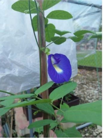
Blue Butterfly pea plant.

Ferdinand A. Dumalagan, Caryl Joy Alvares, and Robert L. Salamasan, from the different institute of the Philippines, wrote a research article aboute, Blue Butterfly Pea: Phytochemicals and E. coli Inhibition, entitled, "Qualitative phytochemical analysis and inhibitory property of Blue Butterfly Pea flower extract against Escherichia coli".This research paper published by the International Journal of Biosciences | IJB, an open access scholarly research journal on Biosciences, under the affiliation of the International Network For Natural Sciences | INNSpub, an open access multidisciplinary research journal publisher.

Abstract

From ancient times, medicinal plants have been utilized to heal a variety of human illnesses. The development of antibiotics as a means of treating diverse bacterial illnesses sparked a revolution in medicine. Several therapeutic plant extracts are known to have antibacterial properties that are applied in the preservation of food and for therapeutic purposes. Blue butterfly pea (Clitoria ternatea) is a plant that is popularly used as medicine and recognized to treat a number of illnesses. Practically, every component of this plant is said to have therapeutic properties. Furthermore, C. ternatea plant has strong antibacterial effect as it contains biological compounds that act as best antimicrobial property against bacteria. Also, phytochemical analysis of C. ternatea flower extract showed the presence of flavonoids, steroids and tannins in which these phytochemical compounds contributed to the antibacterial ability of the flower extract. On the other hand, C. ternatea flower extract has a heavy amount of flavonoids and these results to a very active capacity to inhibit the growth of the Escherichia coli. Hence, flavonoids are reported that it has the capability against infectious degenerative disease, viral and bacterial. Certainly, this medicinal plant may be used for an alternative medicine considering the fact that this is harmless, low-cost production and its flower can be eaten raw. 

Submit your article to  IJB Journal

 Read more:  Bilaspur's Wild Mushrooms: Edible and Medicinal Diversity | InformativeBD

Introduction

Human illnesses are treated using medicinal plants and have been for a very long time. The development of antibiotics as a means of treating diverse bacterial illnesses sparked a revolution in medicine. However, their indiscriminate usage has caused an alarming growth in the number of microorganisms that are resistant to antibiotics, giving rise to multiresistant strains, which has raised concerns around the world (Shariff, 2001).

Clitoria ternatea L. is a species of Clitoria under Fabaceae family is often utilized as a traditional plant called as blue butterfly pea. It is Ayurvedic medicinal plant as a memory booster, stress reliever, and calming, antidepressant, anticonvulsant, and anxiolytic also a sedative (Ramkissoon et al. (2013). More so, C. ternatea has been utilized for a number of medical conditions. Uses for its roots include cure heartburn, bloating, constipation, fever, arthritic pain, sore throat, while its seeds are utilized as a treatment for eye and skin conditions, to treat colic and swollen joints with a laxative. The conventional Cuban culture employs either a single root decoction or paired with flowers to induce menstruation and encourage as well as to treat liver and intestinal problems, uterine contractions problems (Mukherjee et al., 2008; Fantz 1991). Furthermore, its flower petal's nutritional value using anthocyanins as a naturally occurring blue coloring in a various cuisine. It is also act as anti-oxidant, antibacterial, anti-inflammatory, and other pharmacological properties that are present in the extract of C. ternatea (Gupta et al., 2010).

Moreover, a large and increasing number of patients in the world used medicinal plants and herbs for health purposes. There are hundreds of biologically active compounds developed from traditional medicinal plants (Inoue, 2018). It is believed that the plant’s medicinal crude extract is more biologically active that isolated compounds due to its synergistic effects (Jana and Shekhawat, 2010).

Based on the presented prior arts that Clitoria ternatea possess medicinal values and clearly benefit the users. This study explores on the screening of the antibacterial activity of phytochemical properties present in blue butterfly pea that cited that contains antibacterial composition against Escherichia coli. Hence, it can be an alternative medicine to cure various diseases that acts as anti-viral, antiinflammatory, anti- allergic, has rich in antioxidants and good treatment for mental illness that contributes to the range of rediscovering essential uses of this plant.

Reference

Alok S, Gupta N, Malik A. 2015. International Journal of Life Sciences Rev 1(1), 1-9.

Chusak C, Thilavech T, Henry CJ, Adisakwattana S. 2018. Acute effect of Clitoria ternatea flower beverage on glycemic response and antioxidant capacity in healthy subjects: A randomized crossover trial. BMC Complementary and Alternative Medicine 18, 1-11. https:// doi.org/ 10.1186/s12906-017-2075-7.

Fantz PR. 1991. Ethnobotany of Clitoria (Leguminosae). Econ Bot 45, 511-520. https:// doi.org /10.1007/BF02930715.

Ferreira D, Gross GG, Hagerman AE, Kolodziej H, Yoshida T. 2008. Tannins and related polyphenols: Perspectives on their chemistry, biology, ecological effects, and human health protection. Phytochemistry 69, 3006-3008.

Gupta GK, Chahal J, Bhatia M. 2010. Old and new aspects. Journal on Pharmacy Research 3, 2610-4.

Haditio SM, Muttaqin Z, Hadi L. 2021. Comparison of Inhibition Zones between Butterfly Pea flower (Clitoria ternatea) and Lemongrass (Cymbopogon citrarus) against Streptococcus mutans and Staphylococcus aureus. Biomedical Journal of Indonesia 7, 374-378. https:// doi.org/ 10.32539/bji.v7i2.313.

Harborne, JB and Williams, CA. 2000. Advances in Flavonoid Research since 1992. Phytochemistry 55, 481-504.

Jamil N, Pa’ee F. 2018. AIP Conference Proceedings 2002, 020044

Kamilla L, Mnsor SM, Ramanathan S, Sasidharan S. 2009. Antimicrobial activity of Clitoria ternatea (L.) extracts. Pharmacologyonline 1:

Ke S. 2018. Recent Progress of Novel Steroid Derivatives and Their Potential Biological Properties. Mini-Rev. Med. Chem. 18, 745-775.

Kumar MN, More D. 2019. Phytochemical analysis and bioactivity of selected medicinal plant of butterfly-pea (Clitoria ternatea L.) used by Kolam tribe Addjoing region of Telangana and Maharashtra states. The Pharma Innovation Journal 8, 417-421.

Lakshan SAT, Jayanath NY, Abeysekera WPKM, Abeysekera WKSM. 2019. A commercial potential blue pea (Clitoria ternatea L.) flower extract incorporated beverage having functional properties. Evidence-based Complementary and Alternative Medicine 2019: e2916914. https://doi.org/10.1155 /2019/2916914

Lijon MB, Meghla NS, Jahedi E, Rahman MA, Hossain I. 2017. Phytochemistry and pharmacological activities of Clitoria ternatea. International Journal of Natural and Social Sciences 4, 1-10.

Mahomoodally MF, Gurib-Fakim A, Subratty AH. 2005. Antimicrobial activities and phytochemical profles of endemic medicinal plants of Mauritius. Pharmaceut Biol 43, 237-242

Mukherjee PK, Kumar V, Kumar NS. 2008. The Ayurvedic medicine Clitoria ternatea-From traditional use to scientific assessment. J Ethnopharmacol 120, 291-301. https://doi.org/10. 1016/j.jep.2008.09.009.

Okwu DE, Josiah C. 2006. Evaluation of the Chemical Composition of Two Nigerian Medicinal Plants in African Journal of Biotechnology 5, 357-361.

Pandey AK. 2007. Anti-staphylococcal activity of a pan-tropical aggressive and obnoxious weed Parihenium histerophorus: an in vitro study. Natl Acad Sci Lett 30, 383-386

Pratap Gowd MJS, Manoj Kumar M, Sai Shankar A, Sujatha B, Sreedevi E. 2012. Evaluation of three medicinal plants for anti-microbial activity. AYU (An International Quarterly Journal of Research in Ayurveda) 33, 423-428. https://doi.org/10.4103/0974-8520.108859.

Ramkissoon JS, Mahomoodally MF, Ahmed N, Subratty AH. 2013. Antioxidant and anti-glycation activities correlates with phenolic composition of tropical medicinal herbs. Asian Pacific J Tropical Medicine 6, 561-9.

Shariff ZM. 2001. Modern Herbal Therapy for Common Ailments. Nature Pharmacy Series Vol. I. 1st Edn, Spectrum Books Limited, Ibandan, Nigeria in Association with Safari Books (Export), Limited UK pp.9-84.

Uma B. 2009. Phytochemical analysis and antimicrobial activity of Clitoria ternatea Linn. against extended spectrum beta lactamase producing enteric and urinary pathogens. Asian J. Pharm. Clin. Res 2, 94-96.

Vu TT, Kim H, Tran VK, Vu HD, Hoang TX, Han JW, et al. 2017. Antibacterial activity of tannins isolated from Sapium baccatum extract and use for control of tomato bacterial wilt. PLoS One 12:e0181499. https://doi.org/10.1371/journal. pone.

Widyarman AS, Sumadi S, Agustin TP. 2018. Antibiofilm effect of Clitoria ternatea flower juice on Porphyromonas gingivalis in vitro. Journal of Indonesian Dental Association 1, 7-12. https://doi. org/10.32793/jida.v1i1.288.

 SourceQualitative phytochemical analysis and inhibitory property of Blue Butterfly Pea flowerextract against Escherichia coli

Bilaspur's Wild Mushrooms: Edible and Medicinal Diversity | InformativeBD

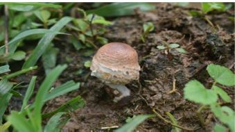
 Different Species of Mushrooms in Bilaspur forest division.

Bhavana Dixit, from the different institute of the india, wrote a research article about, Bilaspur's Wild Mushrooms: Edible and Medicinal Diversity,entitled, "Diversity of edible and medicinal wild mushrooms of Bilaspur District of Chhattisgarh in Central India".This rsearch paper published by the Journal of Biodiversity and Environmental Sciences | JBES. an open access scholarly research journal on Biodiversity, under the affiliation of the International Network For Natural Sciences | INNSpub.an open access multidisciplinary research journal publisher.

Abstract

India is a tropical country with a wide range of climatic conditions; it is a natural habitat for a large range of wild mushrooms. Chhattisgarh, which is the central part of India, has Tropical Forests providing ideal growing conditions for diverse wild mushrooms flora including many edible and therapeutic fungi. The present study explores the biodiversity of naturally growing wild edible and medicinal fungi from the different forest-associated tribal/rural areas in Bilaspur. A total of 15 species of wild edible mushrooms including 8 fungi having therapeutic properties were collected and identified. The identified species were found saprophytic and mycorrhizal. Moreover, the majority of popular edible mushroom species were recorded during either the early or the late rainy season. Therefore, the present study generated a database on mushrooms diversity in the Bilaspur District of Chhattisgarh that will help for its sustainable management.

Submit your article to JBES Journal

Read moreTilapia By-products:Characterizing Fish Protein Hydrolysate | InformativeBD

Introduction

Wild edible fungi have been collected and consumed by people for thousands of years but in recent decades, there has been a surge in global interest in the utilization of wild edible fungi (FAO 2004). Because they are a valuable medicinal, socioeconomic resource, and food safety and, security in many parts of the globe. Wild edible fungi are considered a healthy food because their mineral content is higher than that of meat or fish and most vegetables (Chan, 1981). Furthermore, fresh edible fungi have roughly twice the protein amount of veggies (Chan, 1981). They have rich nutritional value with high content of vitamins, protein and minerals, fibers, trace elements, and contain no or low calories cholesterol (Agahar- Murugkar and Subbulakshmi2005; Wani et al., 2010; Okoro I.O. et al., 2012). Moreover, many researchers have conducted nutritional analyses of various mushroom species, finding that they are high in protein (30–48%), carbohydrate (125–40%), fat (1– 4%), ash (7–17%), and fiber (16% - 20% ), etc. (Pushpa & Purushothama, 2010; Manikandan, 2011). Apart from their use as food, a variety of edible and non-edible fungi have been utilized for therapeutic purposes (Wasser and Weis, 1999; Hobbs, 1995), as they contain a good amount of secondary metabolites and have antioxidant, anticancer, anti-mutagenic, antimicrobial, and antiradical properties (Barros L et al., 2007), that may help to prevent or lessen the risk of cancer, heart disease, diabetes, and viral infections (Oei, 1991). Moreover, the presence or absence of mushroom species can be used to determine whether an ecosystem is degrading or developing, they also play a crucial role in nutrient reprocessing, plant growth, and establishment in forests (Tapwal et al., 2013). More than 2,000 fungi have been identified as producing edible sporocarps around the world (Boa, 2004), and 283 edible species from India (Purkayastha and Chandra, 1985; Adhikari, 2000) among them have been cultivated.

Diversity of edible and medicinal wild mushrooms of Bilaspur District of Chhattisgarh in Central India

The consumption of fleshy fungi has expanded in many nations in recent years, making it necessary to investigate the treasure trove of wild mushrooms. India is a tropical country with a wide range of climatic conditions; it is a natural habitat for a large range of wild mushrooms. Chhattisgarh, which is the central part of India, has Tropical Forests providing ideal growing conditions for diverse wild mushroom flora including many edible and therapeutic fungi. The forests of this state have a high reservoir of unexplored macro-fungal wealth, as the knowledge about various edible and medicinal fungi in different tribal/rural areas has not received significant attention. Therefore, the present study has undertaken to explore the biodiversity of naturally growing wild edible and medicinal fungi from the different forest-associated tribal/rural areas in Bilaspur. This paper reports the wild edible and medicinal mushrooms diversity from Bilaspur district of Chhattisgarh, India

Reference

Adhikari MK. 2000. Mushrooms of Nepal. Kathmandu: P. U. Printers.

Agrahar-Murugkar D, Subbulakshmi GJFC. 2005. Nutritional value of edible wild mushrooms collected from the Khasi hills of Meghalaya. Food Chemistry 89(4), 599-603.

Barros L, Ferreira MJ, Queiros B, Ferreira IC, Baptista P. 2007. Total phenols, ascorbic acid, β-carotene and lycopene in Portuguese wild edible mushrooms and their antioxidant activities. Food Chemistry 103(2), 413-419.

Boa ER. 2004. Wild edible fungi: A global overview of their use and importance to people.

Chakraborty I, Mondal S, Pramanik M, Rout D, Islam SS. 2004. Structural investigation of a water-soluble glucan from an edible mushroom, Astraeus hygrometricus. Carbohydrate Res. 339, 2249- 2254.

Chan HKM. 1981. Consumption of edible mushrooms in Hong Kong. Mushroom Newsletter for the tropics 1(4), 5-10.

Chandrawati SP, Kumar N, Tripathi NN. 2014. A macrofungal wealth of Kusumhi forest of Gorakhpur, UP, India. American International Journal of Research in Formal, Applied, and Natural Sciences 5(1), 71-75.

Das K. 2010. Diversity and conservation of wild mushrooms in Sikkim with special reference to Barsey Rhododendron Sanctuary. NeBIO 1(2), 1-13.

FAO. 2004. Wild Edible Fungi: A Global Overview of Their Use and Importance to People. Non-Wood Forest Products 17. Rome.

Hobbs CH. 1995. Medicinal mushrooms: exploitation of tradition, healing, and culture. Botanica Press. Santacruz pp. 219-251

Manikandan K. 2011. Nutritional and Medicinal Values of Mushrooms. In M. Singh, B. Vijay, S. Kamal, & G. C. Wakchaure (Eds.), Mushrooms Cultivation, Marketing and Consumption 11-14. Solan: Directorate of Mushroom Research.

Oei P. 1991. Manual on mushroom cultivation: techniques, species, and opportunities for commercial applications in developing countries.

Okoro IO, Achuba FI. 2012. Proximate and mineral analysis of some wild edible mushroom. African Journal of Biotechnology 11(30), 7720-7724.

Purkayastha RP, Chandra A. 1985. Manual of Indian Edible Mushrooms. New Delhi, India: Jagendra Book Agency.

Pushpa H, Purushothoma KB. 2010. Nutritional Analysis of Wild and Cultivated Edible Medicinal Mushrooms. World Journal of Dairy and Food Sciences 5, 140-144.

Singha K, Sahoo S, Roy A, Banerjee A, Mondal KC, Pati BR. 2020. Contributions Of Wild Mushrooms In Livelihood Management Of Ethnic. IJPSR 3160-3171.

Sun S. 2014. One kind of traditional Chinese medicine composition for treating diabetes. China patent application CN103550660A.

Tapwal A, Kumar R, Pandey S. 2013. Diversity and frequency of macrofungi associated with wet evergreen tropical forest in Assam, India. Biodiversitas 14, 73-78.

Venkatachalapathi A, Paulsamy S. 2016. Exploration of wild medicinal mushroom species in Walayar valley, the Southern Western Ghats of Coimbatore District Tamil Nadu. Mycosphere 7(2), 118-130.

Wani BA, Bodha RH, Wani AH. 2010. Nutritional and medicinal importance of mushrooms. Journal of Medicinal Plants Research 4(24), 2598-2604.

Wasser SP, Weis AL. 1999. General description of the most important medicinal higher Basidiomycetes mushrooms. Int. Med. Mush 1, 351-370.

Watanabe T. 1937. Pictorial atlas of soil and seed fungi. tsukuba, Ibaraki, Japan: CRC Press Taylor & Francis group an informa business.

SourceDiversity of edible and medicinal wild mushrooms of Bilaspur District of Chhattisgarh in Central India

Tilapia By-products: Characterizing Fish Protein Hydrolysate | InformativeBD

Oven-dried tilapia protein hydrolysate (photo by MJ Rafael).

Mark Joseph R. Rafael, and Ravelina R. Velasco, from the different institute of the philippines, wrote a research article about, Tilapia By-products: Characterizing Fish Protein Hydrolysate, entitled, "Characterization of Fish protein Hydrolysate from Tilapia by-products using acid and enzymatic hydrolysis".This research paper published by the International Journal of Biosciences | IJB. an open access scholarly research journal on Biosciences, under the affiliation of the International Network For Natural Sciences | INNSpub.an open access multidisciplinary research journal publisher.

Abstract

Waste management has been a significant problem in the fish processing industry due to environmental and public health impacts. Food products can be developed from the by-products of the aquaculture industry. This study extracted and characterized fish protein hydrolysate (FPH) from tilapia by-products (viscera). It was produced by enzymatic and acid hydrolysis. The degree of hydrolysis (DH), protein pattern, solubility, emulsifying, and foaming properties of the FPH were determined. The yield of the fish protein hydrolysate increased with increasing concentration for acid hydrolysis. Decreasing total protein was observed with the use of increasing HCl concentration. The DH ranged from 12.79-13.95%. The molecular weight distribution of fish protein hydrolysate using acid and enzymatic hydrolysis was analyzed by SDS-PAGE. Limited hydrolysis formed larger peptides which led to improved emulsification and foaming properties of the fish protein hydrolysate. Tilapia intestine crude enzyme hydrolysis produced FPH with higher solubility in water than using acid solutions. The optimum concentration for acid hydrolysis to produce FPH with high emulsifying activity index was found to be 4M acid solution. The Foaming stability for both the acid and enzymatic hydrolysis were low ranging from 9.17% 10.83%. Based on their characteristics and quality, fish protein hydrolysate extracted using acid and enzymatic hydrolysis were within the criteria that can be used as a value-added product in nutraceutical supplements such as sources of small peptides and amino acids in dietetic foods. The improved solubility, emulsifying and foaming capacities of tilapia protein hydrolysate warrant its application in formulated food systems. 

Submit your article to IJB Journal

Read more : Halophilic Mycoflora:Exploring Coastal Diversity in India | InformativeBD

Introduction

Tilapia are prepared by bleeding, gutting, beheading, filleting, skinning, and trimming before being bought by consumers. The potential use of fish by-products should be considered. Increasing focus on the utilization of fisheries by-products in product development and value addition can be explained through waste management efforts and characterization of the raw materials as a potential food protein source and functional foods. Several food products could be obtained from the wastes of the aquaculture by-products industry. 

Fish protein hydrolysates are products of hydrolysis reaction by breaking the peptide bonds in proteins resulting in shorter peptides or amino acids which are easier for animals to absorb. Extraction of proteins from by-products and conversion to high value products, such as bioactive peptides is a very promising alternative. Bioactive peptide production from fish by-products has received growing attention due to their physiological activities as antioxidant and antihypertensive suitable for healthcare and nutraceutical applications (He et al., 2013; Je et al., 2005; Jung et al., 2006). 

The considerable volume of tilapia produced in the country, aside from the significant requirement for processing before final sale generates a large amount of solid waste or residues and by-products, which account for up to 70% of the total fish weight. These so-called wastes composed of the head, carcass, bones, skin, fins and viscera of tilapia are traditionally considered of low economic value and are disposed in land-based waste disposal system or at sea. Moreover, a large amount of fish is also being discarded each year due to fish kill and disease outbreaks. If not properly discarded or used, they can be an important environmental contamination source since the release of these organic wastes might significantly change the community structure and biodiversity of the benthic assemblages(Caruso, 2015). It is estimated that 32 million tons of waste are produced from the total fish capture and are not used as food (Kristinsson & Rasco, 2000). One of the important waste reduction strategies for the industry is the recovery of marketable by‐products from fish wastes (Arvanitoyannis & Kassaveti, 2008). The study was conducted to produce and characterize fish protein hydrolysate from tilapia by-products.

Reference

Abdul-Hamid A, Bakar J, Bee GH. 2002. Nutritional quality of spray dried protein hydrolysate from Black Tilapia (Oreochromis mossambicus). Food Chemistry 78(1), 69-74. https://doi.org/ 10.1016/S0308-8146(01)00380-6

Arvanitoyannis IS, Kassaveti A. 2008. Fish industry waste: Treatments, environmental impacts, current and potential uses. International Journal of Food Science & Technology 43(4), 726-745. https://doi.org/10.1111/j.1365-2621.2006.01513.x

Bhaskar N, Mahendrakar NS. 2008. Protein hydrolysate from visceral waste proteins of Catla (Catla catla): Optimization of hydrolysis conditions for a commercial neutral protease. Bioresource Technology 99(10), 4105-4111. https://doi.org /10.1016  /j.biortech.2007.09.006

Caruso G. 2015. Fishery Wastes and By-products: A Resource to Be Valorised. Journal of Fisheries Sciences 5.

Cheison SC, Zhang SB, Wang Z, Xu SY. 2009. Comparison of a modified spectrophotometric and the pH-stat methods for determination of the degree of hydrolysis of whey proteins hydrolysed in a tangential-flow filter membrane reactor. Food Research International 42(1), 91-97. https://doi.org/ 10.1016/j.foodres.2008.09.003

El-Beltagy AE, El-Adawy TA, Rahma EH, El-Bedawey AA. 2004. Purification and characterization of an acidic protease from the viscera of bolti fish (Tilapia nilotica). Food Chemistry 86(1), 33-39. https://doi.org/10.1016 /j.foodchem. 2003.08.

He S, Franco C, Zhang W. 2013. Functions, applications and production of protein hydrolysates from fish processing co-products (FPCP). Food Research International 50(1), 289-297. https:// doi.org /10.1016/j.foodres.2012.10.031

Hoyle NT, Merritt JH. 1994. Quality of Fish Protein Hydrolysates from Herring (Clupea harengus). Journal of Food Science 59(1), 76-79. https://doi.org/10.1111/j.1365-2621.1994.tb06901.x

Je J, Park P, Kim S. 2005. Antioxidant activity of a peptide isolated from Alaska pollack (Theragra chalcogramma) frame protein hydrolysate. Food Research International (Ottawa, Ont.), 38(1), 45-50. AGRICOLA. https://doi.org/10.1016 /j.foodres.2004.

Jung WK, Mendis E, Je JY, Park PJ, Son BW, Kim HC, Choi YK, Kim SK. 2006. Angiotensin I-converting enzyme inhibitory peptide from yellowfin sole (Limanda aspera) frame protein and its antihypertensive effect in spontaneously hypertensive rats. Food Chemistry 94(1), 26-32. https://doi.org /10.1016/j.foodchem.2004.09.048

Kim SK, Park PJ, Byun HG, Je JY, Moon SH, Kim SH. 2003. Recovery of Fish Bone from Hoki (Johnius belengeri) Frame using A Proteolytic Enzyme Isolated from Mackerel Intestine. Journal of Food Biochemistry 27(3), 255-266. https://doi.org/ 10.1111 /j.1745-4514.2003.tb00280.x

Klompong V, Benjakul S, Kantachote D, Shahidi F. 2007. Antioxidative activity and functional properties of protein hydrolysate of yellow stripe trevally (Selaroides leptolepis) as influenced by the degree of hydrolysis and enzyme type. Food Chemistry 102(4), 1317-1327.

Kristinsson HG, Rasco BA. 2000. Fish Protein Hydrolysates: Production, Biochemical, and Functional Properties. Critical Reviews in Food Science and Nutrition 40(1), 43-81. https://doi.org /10.1080/10408690091189266

Navarrete del Toro MA, García-Carreño FL. 2003. Evaluation of the Progress of Protein Hydrolysis. Current Protocols in Food Analytical Chemistry 10(1), B2.2.1-B2.2.14. https://doi.org /10.1002 /0471142913.fab0202s10

Ovissipour M, Abedian A, Motamedzadegan A, Rasco B, Safari R, Shahiri H. 2009. The effect of enzymatic hydrolysis time and temperature on the properties of protein hydrolysates from Persian sturgeon (Acipenser persicus) viscera. Food Chemistry 115(1), 238-242. https://doi.org/10.1016 /j.foodchem.2008.12.013

Shahidi F. 2007. Maximising the Value of Marine By-products. Cambridge: Woodhead. http://www. crcnetbase.com /isbn/9781439824542

Silva JFX, Ribeiro K, Silva JF, Cahú TB, Bezerra RS. 2014. Utilization of tilapia processing waste for the production of fish protein hydrolysate. Animal Feed Science and Technology 196, 96-106. https://doi.org/10.1016/j.anifeedsci.2014.06.010

Šližyte R, Daukšas E, Falch E, Storrø I, Rustad T. 2005. Yield and composition of different fractions obtained after enzymatic hydrolysis of cod (Gadus morhua) by-products. Process Biochemistry 40(3-4), 1415-1424. https://doi.org/10.1016 /j.procbio.2004.

Stoyanov J, Hobman J, Brown N. 2001. CueR (YbbI) of Escherichia coli is a MerR family regulator controlling expression of the copper exporter CopA. Molecular Microbiology. https://onlinelibrary.

Wisuthiphaet N, Kongruang S, Chamcheun C. 2015. Production of Fish Protein Hydrolysates by Acid and Enzymatic Hydrolysis. Journal of Medical and Bioengineering, 4(6), 466-470. https://doi.org /10.12720/jomb.4.6.466-470

Yang H, Xue Y, Liu J, Song S, Zhang L, Song Q, Tian L, He X, He S, Zhu H. 2019. Hydrolysis Process Optimization and Functional Characterization of Yak Skin Gelatin Hydrolysates Journal of Chemistry; Hindawi. https://doi.org/10.1155/2019/9105605

SourceCharacterization of Fish protein Hydrolysate from Tilapia by-products using acid and enzymatichydrolysis