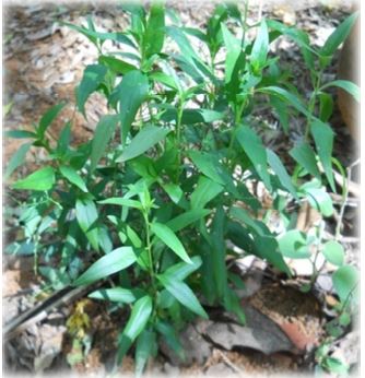
M. Revathy, C. Aswathy,
S. Amutha, E. Amutha, T. Madhumitha, E. Pushpalakshmi, R. Venkateshwari, S.
Rajaduraipandian, and G. Annadurai from the different institute of
the India. wrote a research article about, Nano Silver from Andrographis
paniculata: Germicidal Power. entitled, "Rapid biosynthesis of highly
stabilized nano silver by Andrographis paniculata leaf extract; A prioritized
medicinal plant and its germicidal activity". This research paper published
by the Journal of Biodiversity and Environmental Sciences | JBES. an
open access scholarly research journal on Biodiversity under the affiliation of
the International Network For Natural Sciences | INNSpub. an open
access multidisciplinary research journal publisher.
Abstract
The present research
explains a rapid and environmental friendly route for the synthesis of highly
stabilized silver nanoparticles using the Andrographis paniculata leafextract. The biosynthesized silver nanoparticles were characterized by using
UV-visible spectrophotometer, Fourier Transform Infrared spectroscopy, X-ray
diffraction, and Scanning electron microscope. The UV- visible spectrum
exhibited surface Plasmon resonance band at 421 nm signifies the presence of
silver nanoparticles in the reaction mixture. The XRD technique revealed the
synthesized nanoparticles were crystalline in nature as well as possess face
centered cubic geometry. FTIR studies were carried out to investigate the
functional groups responsible for the silver nanoparticle reduction in the
range 4000 – 400cm-1. EDAX analysis displayed the elemental composition in the
sample. SEM analysis confirmed the synthesized particles are polydispersed in
nature. The inhibition zone gradually increased with increase in silver
nanoparticle concentration. Further the silver nanoparticle exhibited an
effective germicidal activity against both Gram positive and Gram negative
organisms by using disc diffusion method. The in-vitro antimicrobial
assay demonstrated the results of maximum inhibition zone with the (36±0.334)
in Lactobacillus sp and minimum zone with (15±0.318) in Streptococcus
sp. Because of the potent antimicrobial activity it might be concluded that silver
nanoparticles were efficiently utilized as an effective antibacterial
compounds.
Read more : Arugula's Viability Under Semiarid Residual Effects | InformativeBD
Introduction
Nanoscience and nanotechnology is a promising area of nanoscale structures and materials appliances typically ranging from 1 – 100 nm and at these range materials physical and chemical characteristics are drastically distinct from those in bulk materials because of quantum effects (Sharma et al., 2009; Matos et al., 2011). Due to their distinctive properties as well as growing use for several applications in nanomedicine, silver nanoparticles have attracted considerable importance among the rising nanoproducts. Silver in the silver nitrate or silver sulfadiazine form has been utilized for the healing of bacterial infections connected along with burns and injuries due to its antimicrobial properties for a long time (Lok et al., 2007). Several physical, chemical and biological techniques have been improved for the silver nanoparticle synthesis and it has a low yield as well as complex to synthesize nanoparticles with a definite size (Malik et al., 2010).
Traditional approaches similar to physical and chemical such as lithography, laser ablation, pyrolysis, chemical vapor deposition, electrodeposition, sol-gel techniques for nanoparticle synthesis appear to be costly and harmful. Additionally the process includes numerous reactants such as sodium borohydride, potassium bitartrate, methoxypolyethylene glycol, hydrazine as well as it involves stabilizing agents sodium dodecyl benzyl sulfate, polyvinyl pyrrolidone to inhibit the metallic nanoparticles agglomeration. Though numerous techniques are obtainable for the nanoparticle synthesis, there is an essential to expand easy, cost effective, and ecofriendly methods. Hence it is vital to find an alternative biological process for metal nanoparticle synthesis with control particle size and shape for several biomedical applications (Gurunathan et al., 2009; Gurunathan et al., 2013). Plant, plant products, algae, fungi, yeast, bacteria and viruses are a huge collection of biological reserves may possibly utilized for the nanoparticle synthesis and the period needed for the absolute reduction is lesser in biological methods. Biosynthesized nanoparticles are quickly available in solution with high stability, density and depend upon the compounds like alkaloids, tannin, steroids, phenol, saponins, and flavonoids in the plant extract we assume whether the proteins, polysaccharides or secondary metabolites are able to reduce the Ag+ to Ag0 state and develop silver nanoparticles (Singhal et al., 2011). Andrographis paniculata is frequently recognized as king of bitter belongs to the family Acanthaceae indigenous to India and Srilanka. The leaves and root were utilized predominantly for therapeutic purposes in traditional ayurvedic and siddha medicine in India and several other countries. Extract of this plant revealed antifungal, antityphoid, antioxidants, anti-snake venom, anti-inflammatory and antipyretic properties. Andrographis paniculata extract contains two major chemical components acquired from the whole plant such as diterpenoids and flavonoids 7, 2’, 3’- tetramethoxyflavonone as well as 5- hydroxy-7, 2’, 3’-trimethoxyflavone which are expected to be responsible for the major bioactivities of this plant (Aliyu et al., 2009; Puri et al., 1996; Tang et al., 1992).
A number of novel antibiotics were exploited in the previous decades; none have recovered action against multidrug resistant bacteria and due to the growing pervasiveness of microbial resistance has made the public health organization a major concern in the recent world (Mohanty et al., 2012). The main objective of the current study was to improve an easy and ecofriendly method for the silver nanoparticle synthesis and characterization by employing Andrographis paniculata. After that aim of this analysis engaged the germicidal activity of green synthesized silver nanoparticles against both Gram positive and Gram negative organisms. Improving silver nanoparticles as an innovative origination of antimicrobial agents could be a desirable and cheaper means to overwhelm the multi-drug resistance difficulties noticed with bacteria.
Reference
Ahmad N, Sharma S. 2012.
Green synthesis of silver nanoparticles using extracts of Ananas comosus. Green
Sustain Chem 2, 141-147.
Aliyu AB, Musa AM,
Sallau MS and Oyewale AO. 2009. Proximate Composition, Mineral Elements
and Anti-nutritional Factors of Anisopus mannii N. E. Br.
(Asclepiadaceae). Trends Appl. Sci. Res 4(1), 68-72.
Bankar A, Joshi B,
Kumar AR, Zinjarde S. 2009. Banana peel extract mediated novel route for
synthesis of silver nanoparticles. Colloid Surf A Physicochem Eng Aspect 368, 58-63.
Bar H, Bhui DK, Gobinda
SP, Sarkar PM, Pyne S, Misra A. 2009. Green synthesis of silver
nanoparticles using seed extract of Jatropha curcas. Physicochem Eng.
Aspects 348, 212-216.
Berger TJ, Spadaro JA,
Chapin SE, Becker RO. 1976. Electrically generated silver ions:
Quantitative effects on bacterial and mammalian cells. Antimicrob Agents
Chemother 9, 357-358.
Dubey SP, Lahtinen M,
Sillanpaa M. 2010. Green synthesis and characterization of silver and gold
nanoparticles using leaf extract of Rosa rugosa. Colloid Surf A
Physicochem Eng Aspect 364, 34-41.
Dwivedi AD, Gopal K. 2010.
Biosynthesis of silver and gold nanoparticles using Chenopodium album leaf
extract. Colloid Surf A Physicochem Eng. Aspect 369, 27-33.
Geethalakshmi E, Sarada
DV. 2010. Synthesis of plant-mediated silver nanoparticles using Trianthema
decandra extract and evaluation of their antimicrobial activities. Int. J.
Eng. Sci. Tech. 2, 970-975.
Gibbins B. 2003.
The antimicrobial benefits of silver and the relevance of micro lattice
technology. J. Inorg Biochem. 101, 291-296.
Gurunathan S, Han JW,
Eppakayala V, Jeyaraj M, Kim JH. 2013. Cytotoxicity of biologically
synthesized silver nanoparticles in MDA-MB-231 human breast cancer cells.
Biomed. Res. Int. 2013, 535-796.
Gurunathan S,
Kalishwaralal K, Vaidyanathan R, Deepak V, Pandian SRK, Muniyandi J, Hariharan
N, Eom SH. 2009. Biosynthesis, purification and characterization of silver
nanoparticles using Escherichia coli. Colloid Surf B 74, 328-335.
Krishnaraj C, Jagan EG,
Rajasekar S, Selvakumar P, Kalaichelvan PT, Mohan N. 2010. Synthesis of
silver nanoparticles using Acalypha indica leaf extracts and its
antibacterial activity against water borne pathogens. Colloids Surf B:
Biointerfaces 76, 50-56.
Lok CN, Hocm, Chen R,
He QY, Yu WY, Sun H, Tam PK, Chiu JF, Checm. 2007. Silver nanoparticles:
Partial oxidation and antibacterial activities. J. Biol Inorg Chem 12, 527-534.
Malik MA, O’Brien P,
Revaprasadu N. 2002. A simple route to the synthesis of core/shell
nanoparticles of chalcogenides. Chem Mater 14, 2004-2010.
Matos RA, Cordeiro TS,
Samad RE, Vieira JrND, Courrol LC. 2011. Green synthesis of stable silver
nanoparticles using Euphorbia milii latex. Colloid Surface A 389, 134-137.
Mohanty S, Mishra S,
Jena P, Jacob B, Sarkar B, Sonawane A. 2012. An investigation on the
antibacterial, cytotoxic and antibiofilm efficacy of starch-stabilized silver
nanoparticles. Nanomed: Nanotechnol Biol. Med. 8, 916-924.
MubarakAli D, Thajuddin
N, Jeganathan K, Gunasekaran M. 2011. Plant extract mediated synthesis of
silver and gold nanoparticles and its antibacterial activity against clinically
isolated pathogens. Coll Surf B 85, 360-365.
Prabhu S, Poulose EK. 2012.
Silver nanoparticles: mechanism of antimicrobial action, synthesis, medical
applications, and toxicity effects. Int Nano Lett 2, 1-10.
Puri A, Saxena R,
Saxena RP, Saxena KC, Srivastava V, Tandon JS. 1996. Immunostimulant
Agents from Andrographis paniculata, J. Natural Prod 56(7), 995-999.
Sathyavathi R, Krishna
MB, Rao SV, Saritha R, Rao DN. 2010. Biosynthesis of Silver Nanoparticles
Using Coriandrum sativum Leaf Extract and Their Application in
Nonlinear Optics, American Scientific Publishers 3(2), 138-143(6).
Sharma VK, Yngard RA,
Lin Y. 2009. Silver nanoparticles: Green synthesis and their antimicrobial
activities. Adv. Colloid Interface Sci 145, 83-96.
Singhal G, Bhavesh R,
Kasariya K, Sharma AR, Singh RP. 2011. Biosynthesis of silver nanoparticles
using Ocimum sanctum (Tulsi) leaf extract and screening its
antimicrobial activity. J Nanoparticle Res. 13, 2981-2988.
Song JY, Kim BS. 2009.
Rapid biological synthesis of silver nanoparticles using plant leaf extract.
Bioprocess Biosyst Eng 32, 79-84.
Tang W, Eisenbrand G. 1992.
Chinese Drugs of Plant Origin, Chemistry, Pharmacology and Use in Traditional
and Modern Medicine, Springer Verlag, Berlin pp. 97-103.
Williams RL, Doherty
PJ, Vince DG, Grashoff GJ and Williams DF. 1989. The biocompatibility of
silver. Crit Rev. Biocompat 5, 221-243.
Zahir AA, Rahuman AA. 2012.
Evaluation of different extracts and synthesized silver nanoparticles from
leaves of Euphorbia prostrata against Haemaphysalis bispinosa and Hippobosca
maculate. Vet. Parasitol 187, 511-520.
Source : Rapid biosynthesis of highly stabilized nano silver by Andrographis paniculata leaf extract; A prioritized medicinal plant and its germicidal activity


%20in%20full.JPG)


0 comments:
Post a Comment